This is one of 14 Profiles on Bacteria and Archaea. It shows some common bacteria that can be isolated from human sources or other environments. Other Profiles on this site that cover bacteria are: |
|||
This site is no longer maintained and has been left for archival purposes
Text and links may be out of date
This is one of 14 Profiles on Bacteria and Archaea. It shows some common bacteria that can be isolated from human sources or other environments. Other Profiles on this site that cover bacteria are: |
|||
| Bacterial
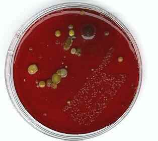
colonies and cell shapes The image below shows a plate of blood agar inoculated by rolling cottonwool swabs over the surface, then it was incubated at 30oC for 6 days. The diagonal swab on the right-hand side was from the surface of the teeth. The diagonal swab on the left was from the sole of a shoe. Note the uniformity of the bacterial colonies from the mouth - an environment of relatively stable temperature, pH etc. and therefore favouring the few species adapted to such conditions. In addition, saliva contains the enzyme lysozyme which degrades the cell wall of Gram-positive bacteria. Compare this uniformity with the diversity of bacterial types from the non-selective environment of the shoe. |
|
|
|
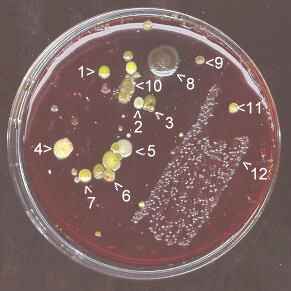
The image above was obtained by placing an open agar plate face-down on a flat-bed scanner with the scanner lid closed. Below we see the same plate scanned with the lid open. The colonies from the mouth (mainly Streptococcus mutans, a primary cause of tooth decay) are embedded in extracellular polysaccharide which helps this bacterium to adhere to the teeth and probably helps to protect the cells from the action of lysozyme (see Exopolysaccharides). |
|
|
|
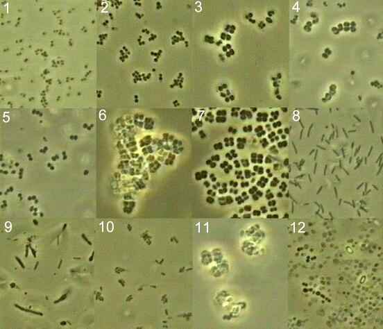
| Samples were taken from the numbered
colonies and examined microscopically, using
phase-contrast optics. The diversity of cell types is
shown below, where all images are reproduced at the same
magnification and in similar optical conditions. Click on any image (labelled 1-12) to see the cells at higher magnification |
|
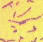
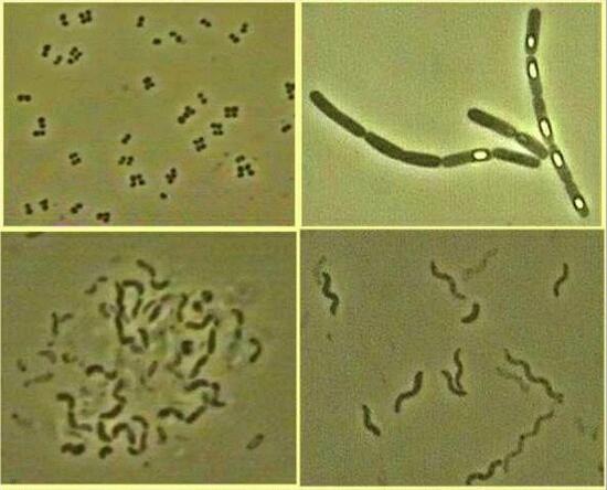
| Bacterial shape The Figure below shows four of the common and characteristic shapes of bacteria: cocci (spherical), rods, vibrio (comma shaped) and spiral. Cocci. The organism shown (top left) is Micrococcus luteus, which forms tetrads due to regular alternation of the plane of cell division. Micrococcus species are commonly associated with the skin. Staphylococcus species also are common on the skin, and S. aureus causes boils or, occasionally, more serious infections. Streptococcus species also are common but typically form chains of cells because they divide in a single plane. Streptococcus mutans commonly grows on tooth enamel and contributes to tooth decay (dental caries). Rods. The organism shown (top right) is Bacillus cereus, a common inhabitant of soil but also found on many food products. Bacillus species produce endospores, seen in various stages of development in the photograph; eventually they are released when the cells lyse. B. cereus causes a relatively mild food poisoning, especially on reheated fried rice in take-away food outlets. The spores can resist destruction during cooking and can then germinate if the cooked food is not refrigerated. Two toxins are formed: one is heat-stable, is produced during sporulation and causing vomiting; the other is heat-labile, is produced during exponential growth and causes diarrhoea. |
|
 |
|
Vibrio. The organism shown (bottom left) is Vibrio alginolyticus, an agar-degrading species found in marine environments. The organisms in the photograph are clustered together in an extracellular polysaccharide (slime) that they synthesized, and many of them are dividing so they are seen in pairs. The best-known species of Vibrio is V. cholerae which causes cholera, a severe diarrhoeal disease resulting from a toxin produced by bacterial growth in the gut. Spiral. The organism shown (bottom right) is a species of Rhodospirillum, one of the purple non-sulphur bacteria (see Winogradsky column). Typically, these bacteria grow in shallow anaerobic organic-rich pools, obtaining energy from light reactions but using organic substances such as acetate for cellular synthesis. [Organisms of this type are termed photoheterotrophs]. However, they can also photosyntheize in anaerobic conditions, obtaining energy from light and fixing carbon dioxide into cellular materials. Rhodospirillum swims in a corkscrew manner, by means of its polar flagella. Other spiral-shaped bacteria include the spirochaetes, such as Treponema pallidum which causes syphilis. However spirochaetes have a different type of motility from that of the common spiral bacteria. |
|
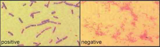
The Gram reaction is named after the Danish physician, Christian Gram, who developed this staining technique in 1884. It involves a series of simple steps.
The method separates bacteria into two types. Gram-positive cells retain the crystal violet-iodine complex and thus appear purple (shown for Bacillus cereus in the left-hand image below). Gram-negative cells are decolourised by the alcohol or acetone treatment, but are then stained with safranin so they appear pink (shown for Pseudomonas aeruginosa in the right-hand image below). Thus, the essential difference between Gram-positive and Gram-negative cells is their ability to retain the crystal violet-iodine complex when treated with a solvent.
|
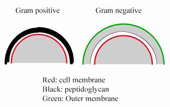
This difference in staining reflects a fundamental difference in the organisation of the bacterial cell wall or "cell envelope", shown in highly simplified form below.
Gram-positive bacteria have a relatively thick wall composed of many layers of the polymer peptidoglycan (sometimes termed murein). The thickness of this wall blocks the escape of the crystal violet-iodine complex when the cells are washed with alcohol or acetone. Gram-negative bacteria have only a thin layer of peptidoglycan, surrounded by a thin outer membrane composed of lipopolysaccharide (LPS). The region between the peptidoglycan and LPS layers is termed the periplasmic space (coloured grey in the figure); it is a fluid or gel-like zone containing many enzymes and nutrient-carrier proteins. The crystal violet-iodine complex is easily lost through the LPS and thin peptidoglycan layer when the cells are treated with a solvent. |
This site is no longer maintained and has been left for archival purposes
Text and links may be out of date