| Proteus vulgaris The genus Proteus is
classified in the enteric bacteria,
together with Escherichia coli, Salmonella,
Shigella, Enterobacter and Serratia.
All these bacteria are small, Gram-negative rods and are facultative
anaerobes: they ferment sugars in anaerobic
conditions but can use a wide range of organic molecules
in aerobic conditions.
Some of the enterics are
major pathogens of humans, but Proteus species
are mainly soil inhabitants, particularly common in
decomposing organic matter. Proteus and the
related genus Providencia can quite frequently
cause urinary tract infections.
|
 |
Proteus has
two interesting and notable features, shown in the images
above. First, the cells are highly motile and often swarm
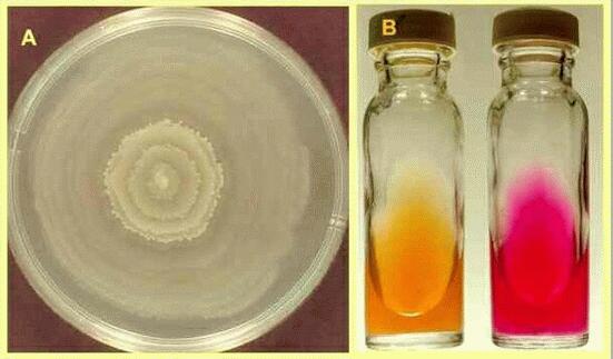
across the surface of agar plates (Figure A).
Swarming gives rise to a very thin film of bacteria on
the agar surface, but swarming periods are interspersed
with periods when the cells stop and undergo a cycle of
growth and division so that the colony has a distinct
zonation, clearly seen in Fig. A.The other notable feature of both Proteus
and Providentia is the ability to degrade urea
to ammonia, by production of the enzyme urease.
This distinguishes them from the other enterics and is
used in a simple diagnostic test (Figure B).
Bacteria isolated from urine samples are inoculated onto
a nutrient agar containing urea and the indicator phenol
red. After overnight incubation, the ammonia produced by Proteus
or Providentia raises the pH and changes the
colour of the medium from yellow to red.
|
Diagnostic
methods in microbiologySimple biochemical tests like the one above
have always been an important aid to identification of
bacteria, because the different bacterial groups and
species have characteristic metabolic activities. A
number of sophisticated tools are now available for
clinical diagnosis. Some are based on monoclonal
antibodies, and others on simple, rapid biochemical
methods.
The API 20E system
shown below has become popular for rapid identification
of members of the Enterobacteriaceae and other
Gram-negative bacteria. The plastic strips consist of 20
small wells containing dehydrated media components (top
row). The bacterium to be tested is suspended in sterile
saline and added to each well, then the strip is
incubated for 16-24 hours and the colour reactions are
noted as either positive or negative. The test results
can be entered into a computer programme to identify the
bacterium.
Four strips inoculated
with four different bacteria are shown in the Figure. In
each case the spectrum of results was different.
|
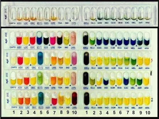 Top row, Proteus vulgaris; second
row, unidentified enteric bacterium; third row, Klebsiella
pneumoniae; bottom row, Vibrio alginolyticus.
|
A few of the tests on these strips are outlined below.On the left-hand side:
- well 1 (marked ONPG)
detects beta-galactosidase activity (yellow for
positive, clear for negative)
- well 3 (LDC) detects
lysine decarboxylase (red positive, yellow
negative)
- well 5 (CIT) detects
citrate utilisation (blue positive)
- well 7 (URE) detects
urease activity (red positive - see the test for Proteus
earlier)
On the right-hand side:
- well 1 (GEL) detects
gelatin liquification (all results were positive
here)
- wells 2-6 detect
fermentation of glucose (GLU), mannitol (MAN),
inositol (INO), sorbitol (SOR) and rhamnose (RHA)
(in each case, yellow positive, blue negative)
GO
TO FULL LIST OF PROFILES?
|