This site is no longer maintained and has been left for archival purposes
Text and links may be out of date
| Some important diseases of humans
transmitted from person to person by inhaled airborne
particles |
|
| Virus diseases (virus type in brackets) |
Bacterial diseases (bacterial name in brackets) |
| Chickenpox (Varicella) | Whooping cough (Bordetella pertussis) |
| Flu (Influenza) | Meningitis (Neisseria species) |
| Measles (Rubeola) | Diphtheria (Corynebacterium diphtheriae) |
| German measles (Rubella) | Pneumonia (Mycoplasma pneumoniae, Streptococcus species) |
| Mumps (Mumps) | Tuberculosis (Mycobacterium tuberculosis) |
| Smallpox (Variola) | |
Several other diseases, below, are acquired by inhaling particles from environmental sources, not directly from an infected person. |
|
| Disease |
Source |
| Psittacosis (Chlamydia psittaci) | Dried, powdery droppings from infected birds (parrots, pigeons, etc.) |
| Legionnaire's disease (Legionella pneumophila) | Droplets from air-conditioning systems, water storage tanks, etc., where the bacterium grows. |
| Acute allergic alveolitis (various fungal and actinomycete spores) | Fungal or actinomycete spores from decomposing organic matter (composts, grain stores, hay, etc.) |
| Aspergillosis (Aspergillus fumigatus, A. flavus, A. niger) | Fungal spores inhaled from decomposing organic matter |
| Histoplasmosis (Histoplasma capsulatum) | Spores of the fungus, in old, weathered bat or bird droppings |
| Coccidioidomycosis (Coccidioides immitis) | Spores in air-blown dust in desert regions (Central, South and North America) where the fungus grows in the soil |
__________________________________________________________________________________________ Psittacosis is a serious disease acquired by handling birds or by inhaling dust from bird faeces. It is caused by the bacterium Chlamydia psittaci, an obligate intracellular parasite. After entering the respiratory tract, the cells are transported to the liver and spleen, multiply there and then invade the lungs, causing inflammation, haemorrhage and pneumonia. Legionnaire's disease is a fairly common form of pneumonia in older or immunocompromised people. It is seldom transmitted directly from person to person. The bacterium is an aquatic rod-shaped species with a temperature optimum of about 36oC, and is a common inhabitant of warm-water systems in buildings. Infection occurs when people inhale aerosol droplets containing the bacteria. Extrinsic allergic alveolitis is a serious hypersensitive response, usually associated with repeated exposure to airborne spores in the work environment. A classic example is the condition termed farmer's lung, caused by exposure to spores of thermophilic actinomycetes. Aspergillosis,
Histoplasmosis and Coccidioidomycosis
are examples of serious fungal infections of humans,
initiated by spores deposited in the alveoli. They can be
life-threatening diseases of immunocompromised people,
when the fungi disseminate from the lungs to major organs
of the body. However, in all cases the infection of
humans is incidental to the fungus, playing no part in
its normal biology. These are fungi that grow naturally
as decomposer organisms in soil, bird faeces or other
organic substrates.
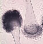
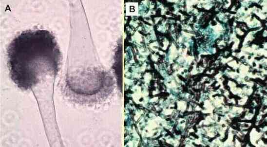
Aspergillus fumigatus. (A) Typical sporing heads of the fungus in laboratory culture. Spores are produced from phialides that arise from the upper part of a club-shaped swelling (vesicle) of an erect hypha (the sporangiophore). [See Thermophilic microorganisms]. (B) Microscopic section of lung tissue, stained to show hyphae of Aspergillus in an air sac. Such a ball of hyphae growing saprotrophically in the lung is termed an aspergilloma. |
| Air-sampling methods Air sampling is used routinely to monitor the populations of airborne particles, and to inform the public about air quality and pollen/spore counts through public broadcasting (weather reports, etc.). It is used by major hospitals to monitor the populations of specific allergenic particles (fungal spores, etc.), so that the causes of patients' allergies can be determined. And it is used in crop pathology for disease-forecasting, so that growers can apply fungicides as and when required. Here we will consider three major types of sampling device for detecting fungal spore loads in air:
|
|
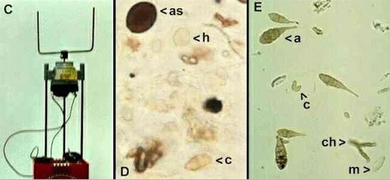
| The Rotorod spore
sampler The rotorod sampler (Figure C below) is a cheap, simple and portable air sampler. It consists of a U-shaped metal rod attached by a spindle to a battery-powered electric motor. The motor causes the upright arms of the metal rod to rotate at high speed. To use the sampler, the upright arms are covered with narrow strips of sticky tape, so that any spores in the air will impact onto the tapes. Then the tapes are removed and examined microscopically to identify the spores and other particles such as pollen grains in the air. Some examples are shown in Figures D and E.
This type of sampler is most effective for trapping relatively large particles (upwards of about 7 micrometres) such as the larger fungal spores and pollen grains. The reason for this is shown in Figure F below.
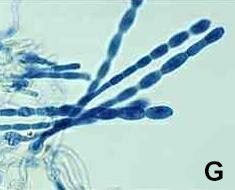
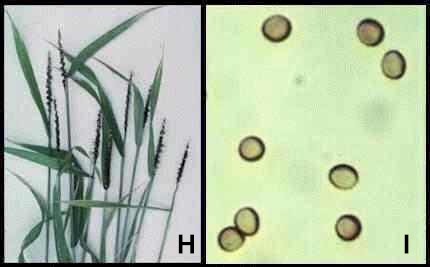
When air is travelling towards an object such as a narrow cylinder, or vice-versa, it is deflected around the object. Any particles in the air will tend to continue along their original trajectory, but their ability to do this (and to impact onto the object) is governed by their momentum (defined as mass x velocity). At any given air speed, the heavier particles are most likely to impact (a in the diagram) whereas smaller (lighter) particles are likely to be deflected round the object. Very high air speeds would be required to impact the smaller particles (b in the diagram), and such air speeds are seldom found in natural conditions. In practice, therefore, fungal spores and other airborne particles can be grouped into two broad categories - those that can impact on surfaces (impactors), and those that are smaller and are only removed from the air by sedimentation in prolonged calm conditions or that are removed from the air by rain. One of the advantages of the rotorod sampler is that it can be used to precisely locate a source of spores of a particular fungus. The famous aerobiologist, PH Gregory, did this in the 1950s by placing rotorod samplers at different positions in a field and "homing in" on a source of spores of the fungus Pithomyces chartarum, which causes a condition known as facial eczema of sheep. Many important pathogens of crop plants have large spores that impact readily onto plant surfaces to initiate infection. Examples include powdery mildew of wheat, Erysiphe graminis (Figure G, with spores about 30 micrometres long) and loose smut of wheat, Ustilago tritici (Figures H, I). This smut disease is characterised by a mass of black spores where the grain would normally be produced. These spores are 8-10 micrometres diameter. (See Biotrophic pathogens).
Further information on the rotorod sampler can be found from the website of a commercial supplier, http://www.multidata.com/samplingtechnologies.html (not on this server). |
|
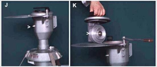
| The Burkard spore trap The Burkard spore sampler acts on the same principle as the rotorod sampler, but is used to give a continuous record of particles in the air over a period of 24 hours or up to 7 days. The apparatus (Figures J, K) consists of an air-sealed drum that contains a clockwork rotating disc (arrowhead in Fig. K) which makes a single revolution in 7 days. The surface of this disc is covered with adhesive tape, to trap spores that impact onto it. When the apparatus is assembled, air is sucked into the drum at high speed through a slit orifice (arrowhead in Fig J) by means of a motor at the base of the apparatus. Any particles in the air impact onto the sticky tape near the slit orifice, giving a record of the particles in the atmosphere at a specific time of day. At the end of a 7-day run, the tape is removed, cut into sections representing hourly or daily periods, then examined microscopically. In this way, it is possible to distinguish clearly between night-released and day-released spores or other particles, and also to relate the types of particle to different weather conditions (e.g. humid or dry periods) while the apparatus was running. The Burkard spore trap is commonly used for continuous monitoring of spore or pollen loads in the air. For example, these traps are commonly sited on hospital roofs, meteorological stations, and other public buildings, and provide public information through TV and radio broadcasts. The principle is exactly the same as in the rotorod sampler because the trapping of particles is based on impaction. The limitations also are the same: only the larger particles with sufficient mass will impact on the tapes at the air speeds generated by this type of sampler.
Figures J-K. The Burkard spore sampling device, shown in assembled form (J) and during preparation (K). |
|
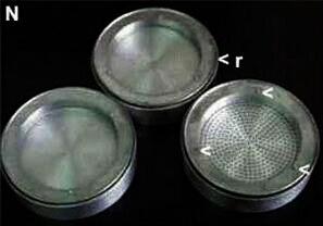
The Anderson sampler The Anderson sampler (Figure L) is an ingenious device for selectively trapping different sizes of particles according to their size (momentum). This sampler consists of a stack of 8 metal sections that fit together with ring seals to form an air-tight cylinder. Each metal section has a perforated base (see Figure N), and the number of perforations is the same in each section, but the size of these perforations is progressively reduced from the top of the column to the bottom. To use this sampler, open agar plates are placed between each metal section, resting on three studs (shown as arrowheads in Figure N).
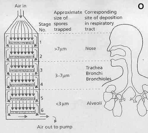
Figures L, M. The Anderson air sampler, shown in a laboratory (L) and mounted with a wind-vane in a field site (M). When fully assembled (with an open agar plate between each unit) an electric motor sucks air from the bottom of the unit, causing spore-laden air to enter at the top (arrowhead in Figure L) and to pass down through the cylinder. The path taken by this air is shown in Figure O, below.
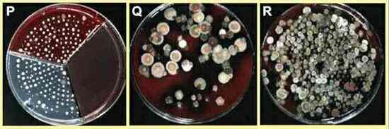
Air sucked in at the top of the column travels at relatively low speed towards the first agar plate, and so only the largest particles impact onto the agar surface. The air then travels round the edge of the agar plate and through the perforations to the second agar plate, and so on. As this process continues down the stack, the same volume of air is forced to travel through successively smaller perforations, and so the air speed is progressively increased. The progressively increased air speed lower down the column raises the momentum of the air-borne particles, so that even the very smallest particles (less than 3 micrometres diameter) can impact onto the lower agar plates. When the sampler has run for 5-15 minutes or more, the metal plates are separated and the Petri dishes are removed for incubation to identify the colonies that develop. Figures P-R (below) show some examples of agar plates from an Anderson sampler. In this case the air sample contained spores from mouldy hay, and the agar plates were incubated at 37oC. Figure P: An agar plate from the bottom level of an Anderson sampler. The colonies are of thermophilic actinomycetes (Micropolyspora faeni or Thermoactinomyces vulgaris) that are common causes of Farmer's lung disease (extrinsic allergic alveolitus). Actinomycete spores are very small (1-2 micrometres) so they commonly enter the lungs. They form dense, slow-growing colonies on agar, and the pattern of colonies seen on this agar plate reflects the pattern of the perforations through which the air had passed. This Figure also shows how a divided (three-sectored) Petri dish can be filled with different agar media to detect different types of organism in the air. Figures Q and R show agar plates from the midlle part of the Anderson sampler, where several species of Aspergillus and Penicillium have developed from spores about 3-5 micrometres diameter. One of the interesting features of the Anderson sampler is that it mimics the deposition of spores (or other ariborne particles) in the human respiratory tract (see Figure O). For example, relatively large fungal spores and pollen grains tend to be trapped on the mucus-covered hairs of our nostrils, where they can cause "hay fever" symptoms in sensitised individuals. Smaller particles are not trapped in the nostrils but instead are carried down into the bronchioles and alveoli. Here the air speed is very low, because the successive branching of the respiratory tract has reduced the air speed to a minimum. But spores of about 2-4 micrometres diameter can settle onto the mucosal surfaces of the alveoli. Some of these spores are important in initiating infections of the lungs. However, it is important to note that the underlying mechanisms of spore deposition in the Anderson sampler are entirely different from those in the human respiratory tract - the Anderson sampler traps spores by impaction, whereas spores are deposited in the human respiratory tract mainly by sedimentation. |
|
The human respiratory tract as an air-sampling device The respiratory tract is highly effective in trapping airborne particles, with sometimes serious consequences for health. The mechanisms involved depend on particle size.
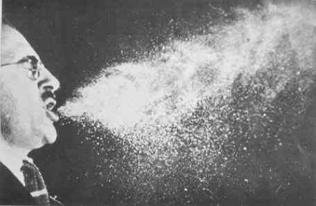
Where do the bacterial and viral pathogens fit into this scheme? The nasopharyngial infections by viruses are associated with large sneeze droplets which impact in the upper airways. Most bacterial diseases also are initiated in the upper airways, when bacteria are carried in large droplets or on "rafts" of skin that impact onto the mucosa. However, infections by Mycobacterium (tuberculosis) and Bacillus anthracis (anthrax) are initiated in the lungs. These are highly virulent pathogens, and even single cells or spores (about 3 micrometres for Bacillus) can initiate infections after deposition in the alveoli. |
|
| Further reading Books: PH Gregory (1973) Microbiology of the Atmosphere. Second Edition. Leonard Hill, Aylesbury J Lacey (1988) Aerial dispersal and the development of microbial communities. pp. 207-237 in: Micro-organisms in Action: Concepts and Applications in Microbial Ecology (Eds JM Lynch & JE Hobbie). Blackwell Scientific Publications, Oxford. Articles: HA Burge (1985) Fungus allergens. Clin. Rev. Allergy 3, 319-329. B Flannigan & JD Miller (1993) Health implications of fungi in indoor environments - an overview. In Health Implications of Fungi in Indoor Environments (eds. R.A. Samson, B. Flannigan, M.E. Flannigan and S. Gravesen), Elsevier, Amsterdam. B Flannigan, EM McCabe & F McGarry (1991) Allergenic and toxigenic micro-organisms in houses. Journal of Applied Bacteriology Symposium Supplement. 70, 61S-73S. Websites: Much useful information can be found from the University of Minnesota Department of Environmental Health and Safety: Fungi in Buildings (not on this server) Other useful links via Aerobiology International (not on this server) |
This site is no longer maintained and has been left for archival purposes
Text and links may be out of date