YeastsYeasts are fungi that grow as single cells, producing daughter cells either by budding (the budding yeasts) or by binary fission (the fission yeasts). They differ from most fungi, which grow as thread-like hyphae. But this distinction is not a fundamental one, because some fungi can alternate between a yeast phase and a hyphal phase, depending on environmental conditions. Such fungi are termed dimorphic (with two shapes) and they include several that cause disease of humans. Here we consider several examples of yeasts and dimorphic fungi:
For further information, see "Dr Fungus" (not on this server) Yeasts grow typically in moist environments where there is a plentiful supply of simple, soluble nutrients such as sugars and amino acids. For this reason they are common on leaf and fruit surfaces, on roots and in various types of food. With few exceptions, they are unable to degrade polymers, such as starch and cellulose which are used by many hyphal fungi. |
||||
|
||||
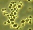
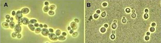
Few of the cellular organelles can be seen by light microscopy, unless they are stained specifically. The only conspicuous organelle seen in Fig. A is the large central vacuole which contributes to cell expansion. S. cerevisiae is a member of the fungal group Ascomycota (the ascus-forming fungi). Cryptococcus albidus (Figure B) is another budding yeast, shown here by phase-contrast microscopy but also with negative staining (the cells are suspended in India ink). Various stages of bud development are seen. The cells are surrounded by a rigid polysaccharide capsule, typical of the genus Cryptococcus, and seen as distinct haloes where the India ink particles have been excluded. Cryptococcus species are common on leaf surfaces. But the most important species from the human standpoint is C. neoformans, a significant pathogen of immunocompromised people, causing the disease termed cryptococcosis. This disease occurs in about 7-8% of AIDS patients in the USA, and a slightly smaller percentage (3-6%) in western Europe. The capsule is a significant virulence determinant of C. neoformans because it helps to prevent the cells from being recognised and engulfed by white blood cells. Additionally, C. neoformans is unique among Cryptococcus species in producing a phenoloxidase. This enzyme acts on phenolic compounds to produce melanin, which might help to protect the cells against the antimicrobial effects of oxidants in host tissues. C. neoformans grows commonly on old "weathered" bird droppings in cities, but does not compete well with bacteria in wet droppings. It infects through the lungs, where it causes a mild or chronic, persistent pneumonia, depending on the person's degree of immunity. Random testing of people for skin reactions to C. neoformans antigens in Britain, Australia and the USA indicates that many people have unknowingly been exposed to the fungus with no serious effect. However, in a small proportion of the population the fungus can disseminate "silently" in the central nervous system, causing fatality. For many years it was assumed that yeast cells inhaled in dried, powdered bird droppings were the source of lung infection. But a sexual stage of the fungus has now been discovered in laboratory culture; it is typical of the fungal group Basidiomycota (which includes the mushroom fungi) but is microscopic, and it leads to the release of small (about 3 micrometre) airborne basidiospores. These are the ideal size for deposition in the lungs (see Airborne Microbes). They are thought to be the main means of infection, but their environmental source is unknown - perhaps a yeast stage growing on vegetation. |
||||
|
||||
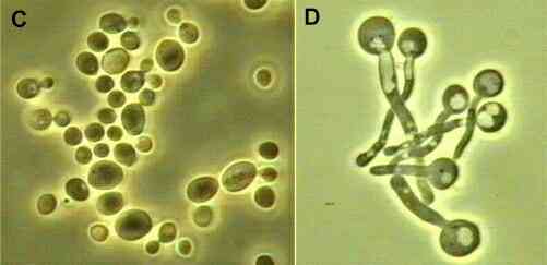
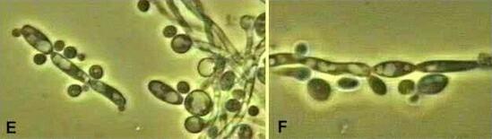
Candida albicans (Figures C, D) Candida albicans is a dimorphic fungus that grows at 37oC. Its normal habitat is the mucosal membranes of humans and other warm-blooded animals, where it grows as a yeast (Fig. C) and causes little or no damage. In fact, it can be isolated from the mucosa of up to 50% of humans - from the mouth, the gut, the vagina or, less often, from the surface of the skin. In some circumstances, however, the same strains of C. albicans that grow as harmless commensals can become pathogenic, invading the mucosa and causing significant damage. This usually happens when a variety of predisposing factors cause the yeast population to multiply, escaping the normal competition from resident bacteria which keep the yeast population in check. Then the yeast cells sprout a hyphal outgrowth (Fig. D) which locally penetrates the mucosal membrane, causing irritation and shedding of the tissues. One of the best examples of this is the disease termed thrush - a white speckling of the tongue and the back of the throat, resembling the speckling on the bird's chest. This is common in newborn babies, perhaps resulting from passage through an infected birth canal. It is also common in AIDS patients and people who have had a prolonged course of antibacterial therapy, reducing the normal resident bacterial population. C. albicans also causes vaginitis - inflammation and invasion of the vaginal mucosa, especially during the third trimester of pregnancy and in women who take the pill. The predisposing factors seem to be hormonal, associated with changes in the balance of cell types in the lining epithelium of the vagina. A similar condition termed stomatitis is common in people who wear dentures. Candida can adhere to denture resin, and high sugar levels in the diet can also increase the adhesion by enhancing the production of a mannoprotein adhesive on the yeast cell surface. Systemic candidosis is a more serious condition, when yeast cells proliferate in the circulatory system. This can occur after invasive surgical techniques, including the insertion of intravenous catheters to which the yeast cells adhere, providing a base from which the cells can bud and be disseminated. All these examples illustrate that C. albicans is a classic opportunistic pathogen, normally kept in check but capable of flaring up in specific, predisposing conditions. It can be identified quite readily from clinical specimens by its ability to sprout hyphae when yeast cells at 37oC are transferred to tubes of horse serum and incubated for 3-5 hours (Fig. D). Only C. albicans and a few related pathogenic Candida species do this. But the fungus has a strong tendency to revert to the yeast phase after only a short period of hyphal growth. Figures E and F show this for horse serum incubated for 24 hours - the hyphae themselves have a beaded appearance, and they give rise to budding yeast cells at the sites where the hyphae of other fungi would form branches. |
||||
|
||||
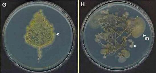
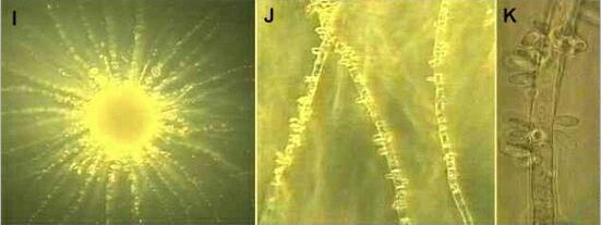
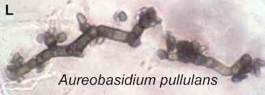
The common occurrence of yeasts on leaf surfaces can be shown by the method in Figure G, where a healthy leaf (in this case from a birch tree) is pressed against the surface of an agar plate for several hours, then removed and the plate is incubated at room temperature. In this example, almost the whole of the leaf print consists of colonies of a single Candida-like dimorphic fungus. The centres of the colonies consist of a mass of yellow-coloured yeast cells, but the fungus is extending across the agar as hyphae at the colony margin (arrowhead). Figures I-K show one of these colonies at increasing magnifications. Instead of branching, the hyphae produce clusters of budding yeast cells at the septa (hyphal cross walls). Older and fallen leaves often have a more diverse fungal community, shown for a fallen oak leaf in Figure H. Again, there are dimorphic fungi (arrowhead) but colonies of Mucor (m) and some darkly pigmented fungi (centre of the leaf print) are also present. The darkly pigmented fungi commonly include Cladosporium species (not shown) and one of the dimorphic "black yeasts", Aureobasidium pullulans (Figure L). |
||||
|
||||
|
||||
|
||||
| Further reading Medical aspects: KJ Kwon-Chung & JE Bennett (1992) Medical Mycology. Lea & Febiger, Philadelphia. NH Georgopapadakou & TJ Walsh (1994) Human mycoses: drugs and targets for emerging pathogens. Science 264, 371-3. |
||||