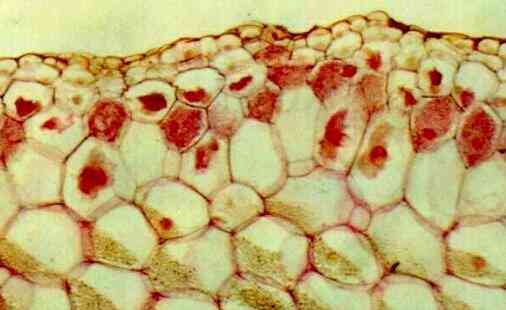
Cross-section of the outer part of the protocorm of an orchid, Neottia, stained to reveal the masses of fungal hyphae (intense red staining) in the outer cortical cells of the protocorm.
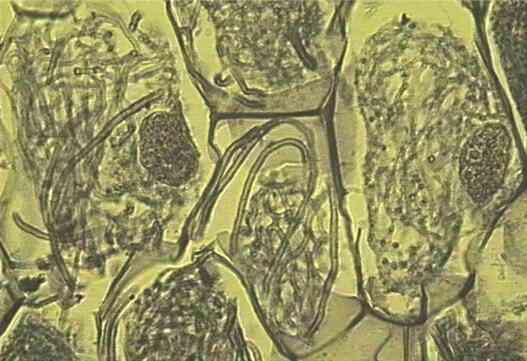
Part of a section at much higher magnification. The cells of the orchid are filled with coils of fungal hyphae but, significantly, the plant cells are still alive and they contain nuclei. The fungal coils will only last a few days or weeks before they are digested (those in the nucleate cell on the right appear to be degenerate) and the process of invasion and digestion will begin again. |