This site is no longer maintained and has been left for archival purposes

Text and links may be out of date

Chytrid zoospores


The Microbial World:
Fungal zoospores II. Chytrids and plasmodiophorids

Produced by Jim Deacon
Institute of Cell and Molecular Biology, The University of Edinburgh

This Profile follows from the introduction given in Fungal Zoospores I. It deals with the zoospores of two further groups of organisms - the chytridiomycota and plasmodiophorids, and their roles as vectors of some important plant 
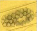
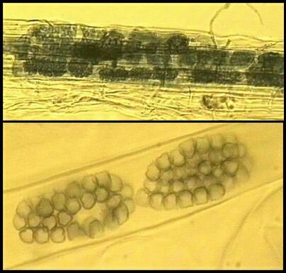
viruses.

1. Chytridiomycota

The chytridiomycota are the only major group of true (chitin-walled) fungi that produce zoospores. These fungi are very common as saprotrophs, facultative parasites and obligate parasites in moist soil and freshwater habitats. They depend on their zoospores for dispersal and site-selection. In fact, this is how the chytridiomycota can be detected - by placing baits such as plant seedlings, pollen grains, insect exoskeletons etc. in water or wet soil, because the zoospores encyst on these materials and then give rise to microscopic colonies (Figure A).


Figure A. Four small colonies of Rhizophlyctis rosea on a piece of Cellophane buried in moist soil for 5 days. The central, globose thallus (body) of each colony has been dislodged, leaving the finely branched rhizoids within the Cellophane.

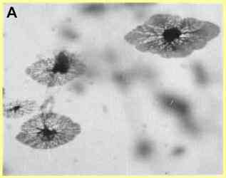
The saprotrophic species include several that degrade polymers. For example, Rhizophlyctis (Karlingia) rosea colonises and degrades cellulose baits in soil, and Chytridium confervae degrades chitin. A specialised group of chytrids grow in the rumen of herbivorous animals, where they degrade cellulose and thus play a primary role in the complex microbial ecology of the rumen. These fungi, such as Neocallimastix frontalis, are unusual because they are obligately anaerobic. They ferment sugars to generate a mixture of formate, acetate, lactate, ethanol, CO2 and H2.

The facultative parasitic species include Catenaria anguillulae, a parasite of nematodes, fluke eggs and several other small organisms (see Figure B). Some other facultative parasites attack the resting spores of plant-pathogenic fungi or mycorrhizal fungi in soil, and perhaps contribute significantly to natural biocontrol systems.


Figure B. Catenaria anguillulae in agar culture. The fungus grows in a hypha-like form, with rhizoids (arrowheads) and chains of swellings (sporangia) in which the zoospores are produced. The generic name Catenaria stems from the latin term "catenulate" (in chains).

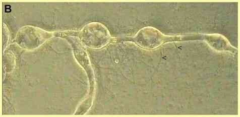
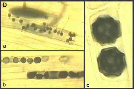
The obligately parasitic species include Olpidium brassicae and other Olpidium species, which are extremely common on roots of wild and crop plants throughout the world. These fungi cannot be grown in culture, away from their hosts. They are symptomless parasites which cause no damage in their own right, but they are important vectors of several soil-borne plant viruses (see later). Stages in the life cycle of these fungi, relevant to their roles as virus vectors, are shown in Figures C and D.


Figure C. Part of a grass root, cleared by treatment with strong alkali then stained with trypan blue to reveal large sporangia (sp) of Olpidium brassicae within the root cells. At maturity the sporangia release zoospores through exit tubes (et). Several zoospore cysts (cy) can be seen on the root.


Figure D. Olpidium brassicae in cleared grass roots. (a) Several zoospore cysts (about 4-5 micrometres) have germinated to produce short tubes that penetrated the root cell wall then released the cyst content as a naked protoplast into the host cell. The protoplasts grow and become multinucleate, then they convert into walled sporangia to release further zoospores. (b, c) Sometimes the host cells contain thick-walled, stellate resting spores (about 15 micrometres), seen in early stages of development (b) and in mature form (c). These spores can persist for several years in soil. They are thought to develop from diploid thalli, when zoospores fuse to form binucleate zoospores which then encyst and infect a host.


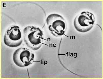
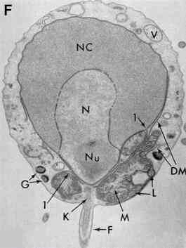
Zoospore ultrastructure


The zoospores of chytridiomycota typically are small and have a single posterior whiplash flagellum (Figures E, F). However, the zoospores of the rumen chytrid Neocallimastix are
larger (about 10 micrometres) and have several flagella.


Figure E. Zoospores of Blastocladiella emersonii viewed by phase-contrast microscopy. The spores are about 2.5 micrometres diameter and contain a prominent nucleus (n), a nuclear cap in which the ribosomes are aggregated (nc), a large mitochondrion (m) near the base of the flagellum (flag), and lipid side bodies (lip). The front end of the cell is a sac-like region containing vacuoles.


Figure F. Transmission electron micrograph of zoospore of Blastocladiella. In addition to the structures mentioned above, the image shows the flagellar motor aparatus (kinetosome, K), Gamma bodies of unknown function (G), and double membranes (DM) that are continuous with the membrane of the nucleus and nuclear cap in regions marked "1".

[Figures E and F supplied by MS Fuller, from Reichle & Fuller, 1967]

Zoospore biology

The basic biology of chytridiomycota zoospores is similar to that of the oomycota, although there are some differences in detail.

Zoospore chemotaxis has been studied for several chytridiomycota, but perhaps the most complete study is for the rumen chytrid Neocallimastix frontalis (Orpin & Bountiff, 1978). In rumen fluid the zoospores showed strong chemotaxis to the awns and other inflorescence parts of barley. In capillary tests on single compounds, chemotaxis was observed towards a range of sugars and sugar derivatives, but not to the common amino acids, purines, pyrimidines and vitamins. By testing the responses to attractants in the presence of high background levels of other compounds (levels that should give saturation binding to receptors) these workers deduced that there are four chemoreceptors on the zoospores:

  • a glucose receptor, which could be saturated by glucose, galactose, xylose, L-sorbose, fucose or 2-deoxy-D-glucose
  • a sucrose receptor, sensitive to sucrose, fructose and raffinose
  • a mannose receptor sensitive to mannose and glucose
  • a sorbitol receptor sensitive to sorbitol and mannitol.

Of all the single compounds tested, the zoospores showed strongest attraction to sucrose, glucose and fructose - the three sugars detected in largest amounts in barley inflorescence tissues - and the strongest attraction was found to a mixture of these three sugars at the concentrations found in awn and inflorescence tissues.

Zoospore settling and encystment has been studied in several chytridiomycota, with some evidence of differential encystment on different materials (Table 1). For example, zoospores of the strongly cellulolytic fungus Rhizophlyctis rosea were induced to encyst after making random contact with pieces of transparent cellulose film or other cellulose sources (cotton threads, etc.) but did not respond to pieces of chitin. Conversely, zoospores of the strongly chitinolytic fungus Chytridium confervae were induced to encyst by chitin but not by cellulose. All possible combinations of responses to these materials were found among the fungi tested (Table 1).

Table 1. Accumulation and encystment of selected zoosporic fungi on pieces of cellulose film and purified crab shell (chitin) in laboratory assays. Data from Mitchell & Deacon, 1986.


Results shown as strong accumulation/encystment or "none" (no difference from controls wihtout the substrates)

  Cellulose Chitin
Allomyces (2 species) Strong Strong
Chytridium confervae None Strong
Rhizophlyctis rosea Strong None
Pythium oligandrum None None
Pythium graminicola Strong Strong
Saprolegnia (3 species) None None

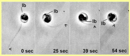
Orientation of encystment in chytridiomycota typically involves a period of amoeboid crawling over a surface (see Catenaria). The anterior sac-like region of these zoospores can send out several pseudopodium-like extensions. Throughout this crawling phase the flagellum is held above the surface, so any cell surface receptors involved in encystment are probably located on the zoospore body, not the flagellum. Finally, in Catenaria the zoospore changes to a rounded shape with the flagellum projecting away from the host surface, and the flagellum is retracted into the zoospore. As shown in Figure G, this retraction is accompanied by rotation of the cell contents.


Figure G. Sequence of photographs from a videotape of an encysting zoospore of Catenaria on a glass surface. In each frame the tip of the flagellum is marked by an arrowhead, and the position of the lipid sidebody complex (lb) is marked..

From frame 1 (0 sec) to frame 4 (after 54 sec) the flagellum is retracted progressively into the cell. This is accompanied by rotation of most or all of the cell contents. The point where the flagellum enters the cell remains in the same position, so it seems that the cell itself remains stationary while the flagellum is reeled in. [From Deacon & Saxena, 1997]


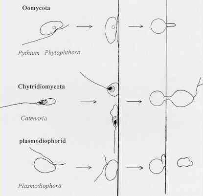
It seems likely that the zoospores of most groups of organisms have a fixed orientation of encystment and germination, as in the oomycota (Figure H). But in both chytridiomycota and plasmodiophorids (e.g. Plasmodiophora brassicae) the zoospores seem to settle with the flagella projecting away from a host surface, based on limited evidence to date. It is not possible to say whether the actual site of germination is pre-determined in chytridiomycota, because the cellular markers change position during flagellar retraction.


Figure H. Comparison of orientation of encystment and cyst germination in three groups of zoosporic fungi. For each group of organisms the diagram shows (left to right) the zoospore, its orientation during encystment, and the cyst germination/ infection events.


2. Plasmodiophorids

The plasmodiophorids are a group of uncertain taxonomic status, sometimes termed the "Plasmodiophoromycota" but they are clearly distinct from fungi. They have many characteristic features, including the fact that they grow as naked protoplasmic stages (plasmodia) in their host cells. The zoospores have two anterior whiplash flagella. Further details can be found in Plasmodiophorid Home Page

All members of this group are obligate parasites of plants, algae or other small organisms, including fungi; but only a few of them are economically important:

Plasmodiophora brassicae, which causes clubroot disease of cruciferous crops

Spongospora subterranea, which causes powdery scab of potato tubers, and a 'special form' (nasturtii) of S. subterranea which causes crookroot disease of watercress

Polymyxa graminis and related Polymyxa species, which are common, symptomless parasites of plant roots but which transmit some important plant viruses.

These organisms depend on zoospores for infection. The zoospore encysts on a host root then germinates to form a pre-penetration swelling termed an adhesorium. From this the protoplast is "injected" into the host cell by a bullet-like mechanism. The protoplast then grows into a multinucleate plasmodium which eventually converts into a sporangium to release further zoospores. As an alternative, the multinucleate plasmodium can convert into numerous thick-walled resting spores (Figure I) which are released when the host cells decay and can survive for many years in soil. They germinate eventually to release zoospores.


Figure I. Grass roots cleared of protoplasm to reveal the presence of clusters of small resting spores of Polymyxa graminis. Top: low-power view of a root. Bottom: two clusters of resting spores in a single root cortical cell.


3. Virus transmission

Four groups of soil-borne plant viruses are vectored by zoospores of the plasmodiophorids (Polymyxa or Spongospora) or Olpidium species (Table 2). No other zoospore type is known to act as a vector, so we must ask why this role is restricted to only a few species. The two important features of these vectors seem to be that:

  1. they are obligate parasites, which infect without killing the host cells;
  2. they have a wall-less, protoplasmic phase within the host.

Infections by Olpidium and Polymyxa species are so common in roots that there is a plentiful supply of zoospore vectors for the viruses specialised to exploit them. Moreover, the viruses will benefit from the host-selection activities of their vectors, although little is known about this behaviour. Added to these points, both Olpidium and the plasmodiophorids can survive for several years as resting spores in soil. As explained below, in many but not all cases, these resting spores can harbour virus particles, making it almost impossible to eradicate viruses from a site once they have been introduced.


Table 2. Major examples of plant viruses that can be vectored by zoospores

Virus name Virus host Vector
 
Tobacco necrosis group (isometric)
Tobacco necrosis virus Many hosts O. brassicae
Cucumber necrosis virus Cucumber Olpidium sp.
Melon necrotic spot virus Melon, cucumber Olpidium radicale
Tobacco stunt type (rigid tubular; double-stranded RNA)
Tobacco stunt virus Nicotiana Olpidium brassicae
Lettuce big vein virus Lettuce O. brassicae
Barley yellow mosaic group (filamentous)
Barley yellow mosaic virus Hordeum spp. Polymyxa graminis
Wheat spindle streak mosaic virus Triticum spp. P. graminis
Oat mosaic virus Avena spp. P. graminis
Wheat yellow mosaic virus Triticum spp. P. graminis
Rice necrotic mosaic virus Oryza spp. P. graminis
Furovirus group (Fungally transmitted rod-shaped viruses; single-stranded RNA)
Soil-borne wheat mosaic virus Triticum spp. Polymyxa graminis
Beet necrotic yellow vein virus Beta spp. Polymyxa betae
Potato mop top virus Solanum spp. Spongospora subterranea
Oat golden stripe virus Avena spp. P. graminis
Peanut clump virus Peanut P. graminis
Broad bean necrosis virus Broad bean P. graminis

From the table above, we see that there are four distinct virus types that can be transmitted by zoospores, two of the virus groups being vectored by Olpidium species, and two by plasmodiophorids (usually Polymyxa species). However, the virus-vector relationships differ quite substantially.

Tobacco necrosis-type viruses

These viruses have a single-stranded RNA genome contained within a single type of isometric particle. This particle shape separates them from all the other zoospore-vectored viruses. But there are also further differences.

  • The tobacco necrosis-type viruses are not wholly dependent on zoosporic vectors. Instead, they can survive in soil and infect roots through wounds.
  • They are carried on the surface of the zoospores, not internally. In fact, they are acquired from soil when the virus particles adhere to the surface of the zoospore or the flagellum. Zoospores can be rid of these virus particles quite easily by addition of virus-specific antisera or by mild acid or alkali treatment.
  • They are not present in the resting spores, because they are not acquired by the fungus within the host plant. So they have no opportunity for long-term survival in the vector. This type of virus-vector relationship has been termed non-persistent.

Tobacco stunt-type viruses

These rod-shaped viruses contain double-stranded RNA, but many of their features remain to be investigated. They are acquired by Olpidium while it is growing in plant cells. Evidently, they are carried internally in the zoospores because they cannot be removed by the treatments that remove tobacco necrosis virus types. They also enter the resting spores and survive in soil, but not for as long as the viruses transmitted by plasmodiophorids, which produce extremely persistent resting spores.

Furovirus group

These viruses are characteristically vectored by plasmodiophorid zoospores. The viruses have a single-stranded RNA genome, divided into two major segments (RNA-1 and RNA-2) that occur in different virus particles. Both particles are required for infection of plants (see Furoviruses). These viruses are acquired by the fungus within the plant cells and are carried internally in both the zoospores and the resting spores, with very long persistence times in soil.

Barley yellow mosaic group

Like the furoviruses, the barley yellow mosaic group have single-stranded RNA divided between two particle types. In terms of virus-vector relationships they are identical to the furoviruses, but they are classified separately as the Bymovirus genus in the family Potyviridae.


Further reading:

Books:

MS Fuller & A Jaworski (1987) Zoosporic Fungi in Teaching and Research. Southeastern publishing Company, Athens, Georgia.

Research articles

JW Deacon & G Saxena (1997) Orientated zoospore attachment and cyst germination in Catenaria anguillulae, a facultative parasite of nematodes. Mycological Research 101, 513-522.

RT Mitchell & JW Deacon (1986) Selective accumulation of zoospores of chytridiomycetes and oomycetes on cellulose and chitin. Transactions of the British Mycological Society 86, 219-223.

CG Orpin & L Bountiff (1978) Zoospore chemotaxis in the rumen phycomycete Neocallimastix frontalis. Journal of General Microbiology 104, 113-122.

RE Reichle & MS Fuller (1967) The fine structure of Blastocladiella emersonii zoospores. American Journal of Botany 54, 81-92.

Websites

Plant viruses Online. A comprehensive listing of viruses, biophysical properties, hosts, etc., and electron micrographs (but very large files with excessive loading times).

Plasmodiophorid Home Page

Zoosporic Fungi Online

GO TO FULL LIST OF PROFILES?

This site is no longer maintained and has been left for archival purposes

Text and links may be out of date

Accessibility Statement