..
MORE IMAGES FROM CHAPTER 14: FUNGI AS PLANT PATHOGENS
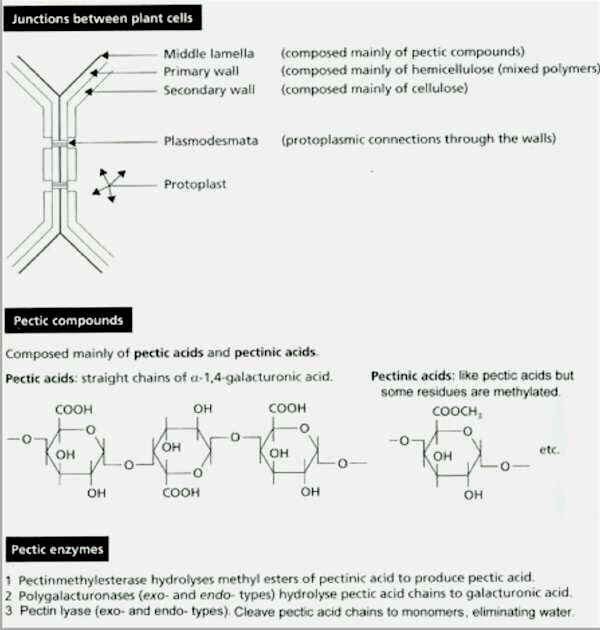
Fig. 14.7. Organisation of the middle lamella (cementing layer) between plant cell walls, and summary of the pectic compounds and pectic enzymes that degrade them. [© Jim Deacon]
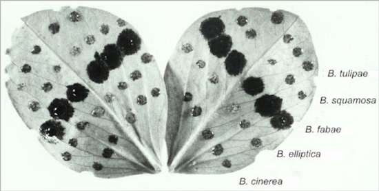
Fig. 14.8. Broad bean leaflets inoculated with water drops containing spores of different Botrytis spp. Only B. fabae is a host-specialised pathogen of broad bean plants; the other Botrytis spp. have different hosts, but B. cinerea is a weak pathogen. [© Photograph courtesy of J. Mansfield.]
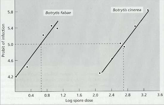
Fig. 14.9. Log dose/probit response plots of infection of broad bean leaves by Botrytis fabae and B. cinerea. Based on Wastie (1960). See text for details. [© Jim Deacon]
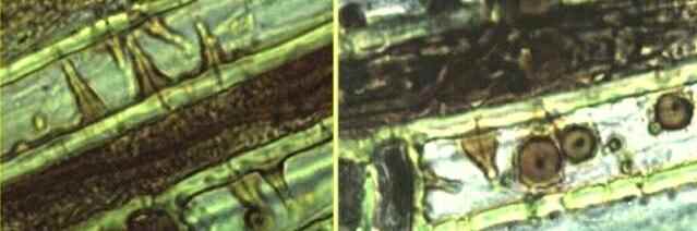
Fig. 14.10. Lignified papillae produced in response to invasion of wheat root cells by the take-all fungus, Gaeumannomyces graminis. Narrow penetration hyphae are seen growing through the cell walls, but they have been stopped by the developing papillae. [© Jim Deacon]
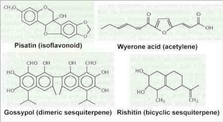
Fig. 14.11. Structures of four phytoalexins: pisatin from peas; wyerone acid from broad bean; gossypol from cotton, and rishitin from potato.
Fig. 14.12. Two examples of detoxification of phytoalexins (at the position marked *)
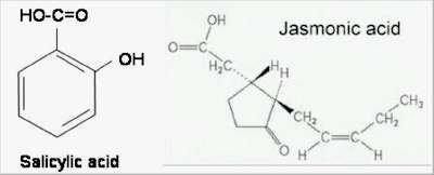
Fig. 14.13. Salicylic acid and jasmonic acid – two key signalling compounds that enhance the resistance of plants to pathogenic attack.
Fig. 14.14. Diagrammatic representation of pathogen spread and occlusion of the xylem vessels in vascular wilt diseases. Spores or yeast-like cells are carried upwards in the water flow and become trapped on the perforated vessel end walls. They germinate, grow through the pores and produce further cells that are carried upwards to the next vessel end wall, and so on. Based on an image by Beckman & Talboys (1981).
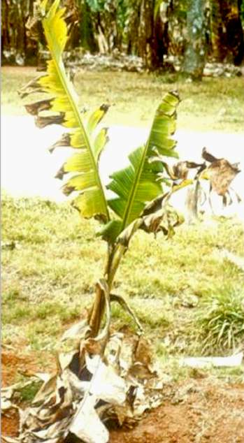
Fig 14.15a. Panama disease of banana plants, caused by Fusarium oxysporum forma specialis (“special form”) cubense. The image shows a young banana plant in an advanced stage of disease; most of the leaves have died and collapsed as a ‘skirt’ around the stem base, and the erect leaves show progressive yellowing and necrosis. [© Jim Deacon]
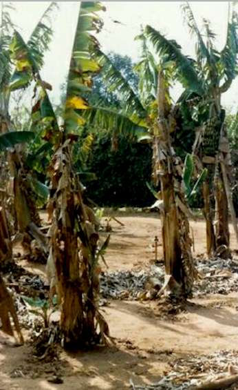
Fig 14.15b Older, fruit-bearing banana plants infected by F. oxysporum , showing extensive collapse of the leaves. [© Jim Deacon] |
|||||||||||