..
MORE IMAGES FROM CHAPTER 14: FUNGI AS PLANT PATHOGENS
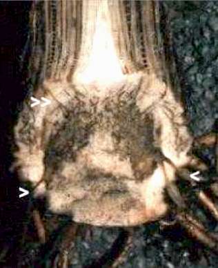
Fig. 14.16a. Section through the base of a diseased banana plant showing heavily discoloured vascular strands (single arrowheads) where the fungus entered the base of the corm from roots, and (double arrowhead) where the infection is spreading up the vascular strands in the leaf sheaths. [© Jim Deacon]
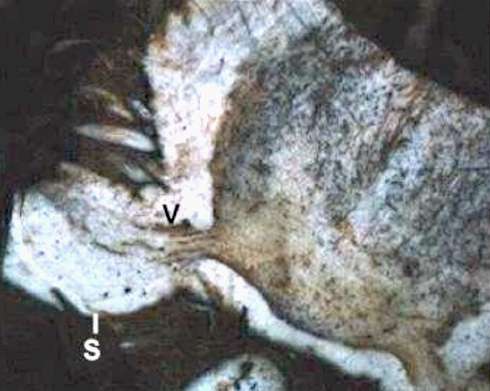
Fig. 14.16b. An older banana corm producing ‘suckers’ (S) that will produce the next banana plant. Vascular disease (V) is already spreading into the new plant. [© Jim Deacon]
Fig. 14.17. Left and Centre: Loose smut of wheat caused by Ustilago nuda. At maturity the flowering spikes produce a mass of black spores in place of the seeds. The image in the centre compares a smutted flowering spike with a normal spike. Right: warty, ornamented spores (smut spores) about 8 µm diameter. [© Jim Deacon]
Fig. 14.18. Percentage of pastures testing positive for Neotyphodium endophytes in a survey conducted by United States Department of Agriculture, 1998. Source: http://w.w.w.aphis.usda.gov/vs/ceah/Equine/eq98endoph.htm
Fig. 14.19. Phytophthora infestans. Left: A sporangiophore, with attached sporangia, emerging from a stoma on the underside of a potato leaf. Right: Two detached sporangia, producing germ-tube outgrowths from near the apical papilla. Note the characteristic broken ‘stalk’ at the base of the sporangia. [© Jim Deacon]
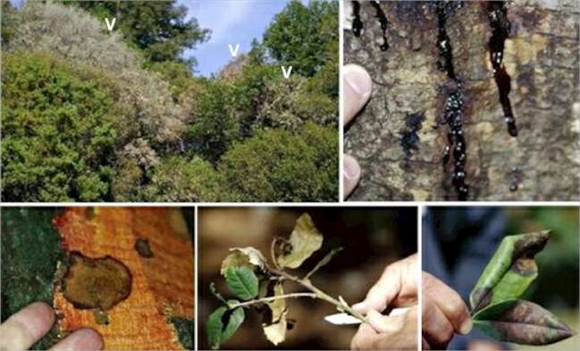
Fig. 14.20. Sudden oak death caused by Phytophthora ramorum. Top left: dead and dying trees of coast live oak (Quercus agrifolia) in the mixed oak community of the coastal fog belt of southwestern USA. Dead or dying trees are indicated. Top right: dark viscous sap is exuded from the bark at the base of a heavily infected coast live oak. Bottom left: removal of the bark reveals the presence of dark zone lines. Bottom centre: terminal die-back and wilting of the shoot tip of tan oak (Lithocarpus densiflorus) is one of the characteristic features of the disease. Bottom right: necrotic, spreading leaf spots are a further symptom of sudden oak death – in this case on leaves of Azalea or Rhododendron. [Images supplied by courtesy of Joseph O’Brien, USDA Forest Service, www.invasive.org; accessed 22 March 2004]
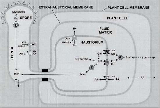
Fig. 14.21. Schematic model for amino acid and sugar uptake and redistribution in rust fungi, based on a diagram in Voegele & Mendgen (2003). Sucrose (Suc) and amino acids (AA) pass between plant cells and through the extrahaustorial membrane, into the fluid-filled matrix that surrounds the haustorium. Invertase (shown as A) cleaves sucrose to glucose (Glc) and fructose (Frc) in the matrix. Then these sugars, and amino acids, cross the haustorial membrane by symport proteins (shown as B) in the membrane. Energy for nutrient uptake is supplied by the H+ gradient generated across the membrane by conversion of ATP to ADP + Pi. Within the haustorium, fructose is converted to mannitol (Man) by the enzyme, major alcohol dehydrogenase (shown as D). Mannitol is then translocated into the fungal hyphae and provides the nutrients for fungal sporulation.
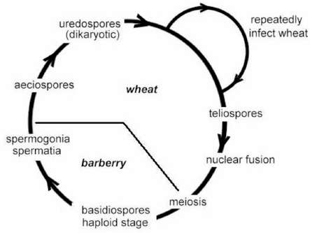
Fig. 14.22. Outline of the disease cycle of Puccinia graminis, which alternates between phases of growth on wheat and on barberry. [© Jim Deacon] |
|||||||||||