..
MORE IMAGES FROM CHAPTER 13: FUNGAL SYMBIOSIS
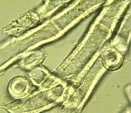
Fig 13.17b Thick-walled, hydrophobic hyphae of the medulla of Peltigera. [© Jim Deacon]
Fig 13.18a Sites of potential nutrient exchange in lichens of the soil crust community of semi-arid desert soils. Three green algal cells (labelled a) are firmly attached to a hypha in the medulla of the lichen Peltula. The arrowhead shows a hyphal projection into an algal cell. [© Jim Deacon]
Fig. 13.18b A cyanobacterial lichen (Collema sp., which is one of the gelatinous lichens). Fungal pegs are closely associated with depressions in the cyanobacterial cells, which are surrounded by a gelatinous sheath. [© Jim Deacon]
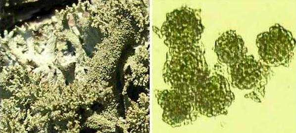
Fig. 13.19. Isidia (left) and soredia (right): two methods of vegetative dispersal of lichens. [© Jim Deacon]
Fig. 13.20a. Part of a thallus of Xanthoria parietina, sectioned through an apothecium. Many asci are seen just beneath the upper surface of the apothecium. [© Jim Deacon]
Fig. 13.20b, Part of a crushed apothecium of Xanthoria parietina, composed of packing hyphae (paraphyses) and asci containing ascospores. [© Jim Deacon]
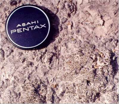
Fig. 13.21. A dried soil crust community of lichens, cyanobacteria and fungal hyphae that form a thin covering which binds the surfaces of semi-arid soils. Such a community would be more than 100 years old and represents a relatively early stage in soil formation. [© Jim Deacon]
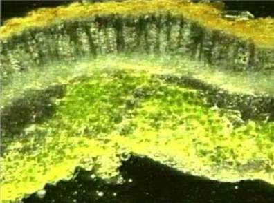
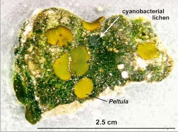
Fig. 13.22. A small fragment of a desert crust community when remoistened, showing several small lichen thalli (Peltula sp.) and one small cyanobacterial lichen (Collema). Most of the soil surface is covered with filaments of the cyanobacterium Scytonema. [© Jim Deacon]
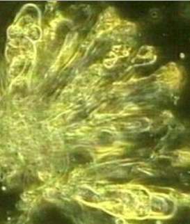
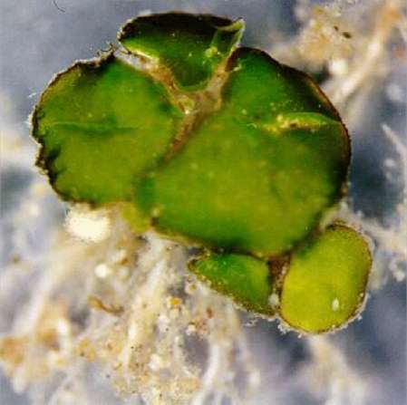
Fig. 13.23a. The desert crust lichen, Peltula, which typically grows as a thallus composed of several squamules. [© Jim Deacon] |
|||||||||||