..
MORE IMAGES FROM CHAPTER
13: FUNGAL SYMBIOSIS
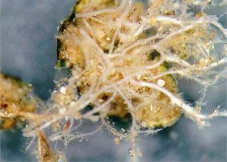
Fig.
13.23b. The same lichen as in Fig. 23a but seen from
below, showing a mass of branched rhizinae that
‘root’ into the desert sand. [© Jim
Deacon]
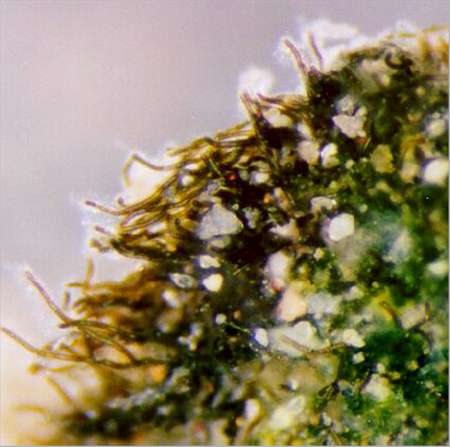
Fig.
13.23c. Part of Fig. 12.22, enlarged to show the
mass of cyanobacterial filaments (Scytonema sp.). [©
Jim Deacon]
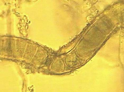
Fig.
13.23d: a single filament of Scytonema encased
in a mucilaginous sheath with soil particles. [©
Jim Deacon]

Fig.
13.24. Bladders of Geosiphon pyriforme growing
on the surface of soil; bar = 1 mm [Courtesy of A.
Schuessler]
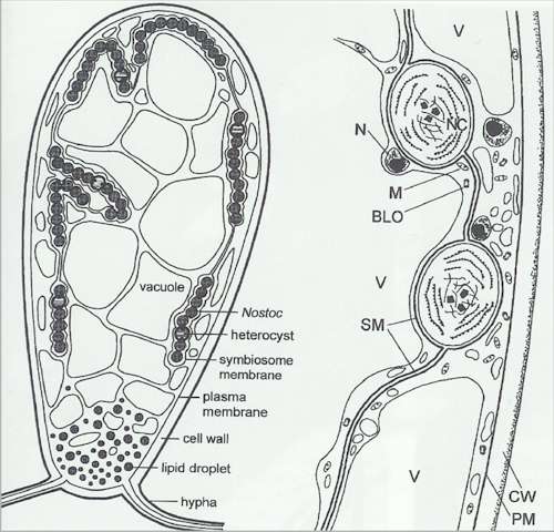
Fig.
13.25. Schematic drawings of the Geosiphon
bladder compartmentation. Left: Cells of Nostoc
are located in membrane-bound symbiosomes towards the
periphery of the fungal cell. Right: detail
showing a bacteria-like organism (BLO), cell wall (CW),
mitochondrion (M), nucleus (N), Nostoc cell (N),
plasma membrane (PM), symbiosome membrane (SM) and
vacuole (V). [Image courtesy of A. Scheussler & M.
Kluge; from Schuessler & Kluge, 2001)
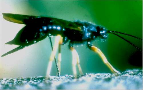
Fig.
13.26. The wood wasp, Sirex noctilio, boring a
hole in a weakened tree to deposit eggs and fungal
spores. [Courtesy of M. P. Coutts, J. E. Dolezal and the
University of Tasmania – see Madden & Coutts,
1979]
GO TO CHAPTER 14 ?
GO TO HOME
PAGE ?
|