..
IMAGES FROM CHAPTER 12:
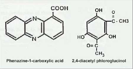
Fig 12.1 Two antibiotics from fluorescent pseudomonads that are widely implicated in control of plant-pathogenic fungi. [© Jim Deacon]
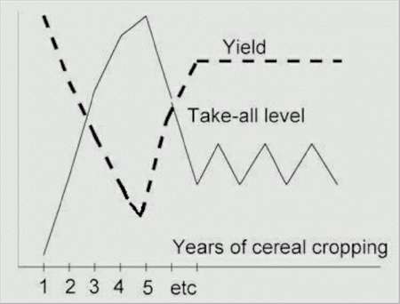
Fig 12.2 Take-all decline when cereals are grown continuously (year after year) in field conditions. [© Jim Deacon]
Fig 12.3 The effects of different population levels of a diacetyl-phloroglucinol producing fluorescent pseudomonad on disease level (or shoot height) of wheat seedlings grown in take-all infested soil. [Reproduced from Raaijmakers & Weller, 1998]
Fig 12.4 Structure of N-hexanoyl-L-homoserine lactone, a quorum-sensing molecule that regulates the synthesis of phenazine antibiotics by fluorescent pseudomonads.
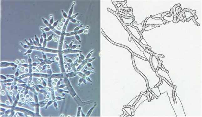
Fig 12.5 Left: Aerial spore-bearing structures of Trichoderma harzianum: the condiophores branch at right angles and produce conidia at the tips of flask-shaped phialides. [Courtesy of Samuels,G.J, Chaverri,P., Farr,D.F. & McCray,E.B. Trichoderma Online, Systematic Botany and Mycology Laboratory, ARS, USDA; from http://nt.ars-grin.gov/taxadescriptions/keys/TrichodermaIndex.cfm] Right: Trichoderma species frequently coil round the hyphae of other fungi (in this case, Rhizoctonia solani) and penetrate or disrupt the host hyphae. [© Jim Deacon]
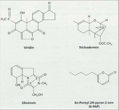
Fig 12.6 Structures of some antibiotics produced by species of Trichoderma
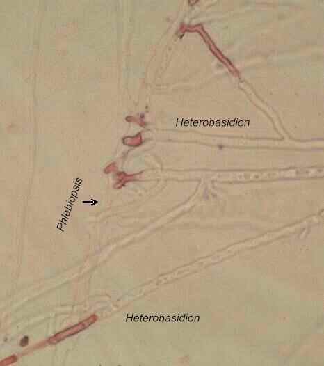
Fig 12.7A. Hyphal interference on an agar plate. Hyphae of Heterobasidion annosum (previously termed Fomes annosus) have been antagonised where they were contacted by single hyphae of Phlebiopsis gigantea (previously termed Peniophora gigantea). The agar plate was then flooded with a dilute solution of neutral red, which was not taken up by the undamaged hyphae but entered the damaged hyphae of Heterobasidion (seen as darker pigmentation). [© Jim Deacon]
Fig 12.7B Hyphal interference of hyphae of Heterobasidion annosum when antagonised by hyphae of Phlebiopsis gigantea. The agar plate was flooded with a dilute solution of neutral red, which was not taken up by the undamaged hyphae but entered the damaged hyphae of Heterobasidion. [© Jim Deacon]
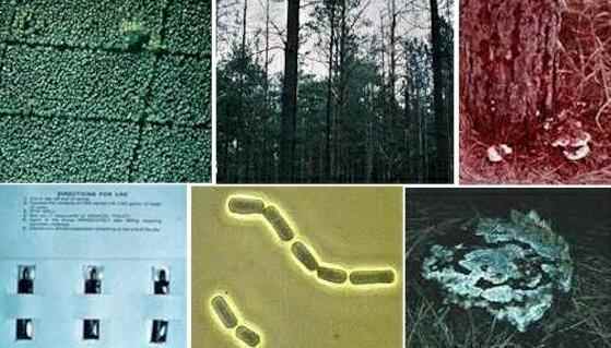
Fig 12.8 Control of pine root rot (Heterobasidion annosum) by Phlebiopsis gigantea. Top left: A pine plantation showing a large disease gap (near the top) caused by H. annosum. Top centre: trees within this gap are spindly and dying. Top right: bracket-shaped fruiting bodies of H. annosum growing from the base of a heavily diseased tree. Lower left: sachets containing spores of P. gigantea, which can be diluted with water and inoculated onto freshly exposed stump surfaces. Lower centre: brick-like spores of P. gigantea are produced abundantly in laboratory culture by fragmentation of the hyphae. Lower right: a pine stump one year after treatment with spore suspension of P. gigantea, showing abundant growth of P. gigantea over the stump surface. [Images courtesy of the late J. Rishbeth]
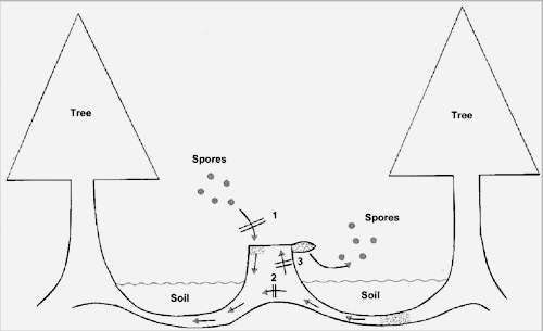
Fig 12.9 Possible mode of action of Phlebiopsis gigantea in controlling Heterobasidion annosum in recently felled pine stumps. 1. P. gigantea is inoculated onto freshly exposed stump surfaces and prevents colonisation of the stumps from airborne basidiospores of H. annosum (shown by the double lines). 2. When P. gigantea has colonised the dying stump and root tissues it prevents the spread of H. annosum from existing foci of infection in the root zone. 3. P. gigantea might also prevent H. annosum from growing up to the stump surface to sporulate. [© Jim Deacon]
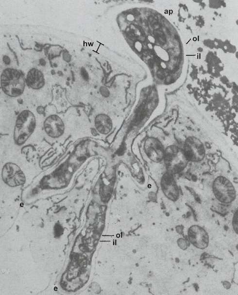
Fig 12.10 Electron micrograph of an appressorium (ap) and a branched haustorium of the mycoparasite Piptocephalis unispora (Zygomycota) in a fungal host, Cokeromyces recurvatus. Hw = host wall; ol and il = outer layer and inner layer of the Piptocephalis wall. The haustorium is surrounded by a continuous membrane (the extrahaustorial membrane, labelled e). [Courtesy of P. Jeffries; from Jeffries & Young, 1976]
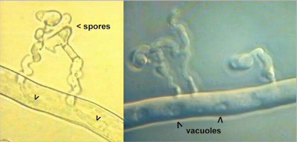
Fig 12.11 Mycoparasitism of Rhizoctonia solani (anastomosis group 4) by Verticillium biguttatum. Left: Spores of V. biguttatum germinate near the host hyphae and grow towards the host in a spiral manner, then penetrate the living hyphae to produce club-shaped haustoria (arrowheads). Right: a similar image, showing that the parasitised host hyphae are still alive; they have intact vacuoles, and videotaped sequences show active protoplasmic streaming. [© P. van den Boogert & Jim Deacon]
Fig 12.12 Antagonism of a hyphal compartment of Fusarium oxysporum by Clonostachys rosea. The damaged compartment has lost its turgor but the adjacent compartments (top left and bottom right) are unaffected. A narrow Fusarium hypha (arrowhead) has regrown into the damaged compartment from a septal pore.[Note: this image has been photo-modified to remove extraneous background features] [© Jim Deacon] |
|||||||||||