CYANOBACTERIA AND THE CRYPTOBIOTIC CRUST
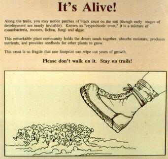
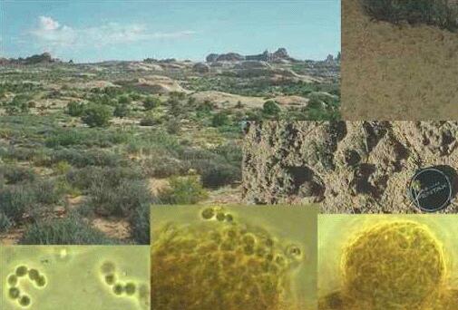
Sign in the Arches National Park, Utah, USA. Probably one of the few signs in the world that says, in effect: Please don't walk on our microorganisms! The image below shows a desert community in the canyonlands area of Utah, USA. This dryland community (top left; comprising saltbush, Pinyon pine, Utah juniper, Indian ricegrass) and all the animals it supports depend on the pioneer role of a microbial community termed the cryptobiotic crust (also known as microbiotic or cryptogamic crust). These microbial communities initially form an inconspicuous grey-brown covering of the sand surface (top right), consisting of fungi, cyanobacteria and lichens, but in later stages of development (after 50 years or more; centre right) the crusts form small "humps" on which mosses grow. The growth of all these pioneer organisms contributes organic matter which aids water retention and paves the way for growth of higher plants. Lichens (see the micrograph, bottom right), which consist of a fungal tissue containing either green algae or cyanobacteria as the photosynthetic partner, play a vital role in colonisation of the bare sand. In this case the lichens contain cyanobacteria (bottom centre and bottom left) which fix atmospheric nitrogen (N2) gas into amino acids and thus progressively enrich the soil with nitrogen for plant growth.
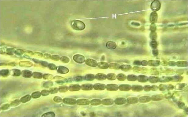
The image below shows a pure culture of the cyanobacterium Nostoc, a common photosynthetic partner of lichens. Nitrogen fixation occurs in special cells termed heterocysts or heterocytes (H) which occur at intervals along the cyanobacterial filaments. This separation of cellular functions is necessary because cyanobacteria have oxygen-evolving photosynthesis but the nitrogen-fixing enzyme, nitrogenase, is unstable in the presence of oxygen. This problem is overcome because the heterocysts contain only part of the photosynthetic apparatus, termed photosystem I, which can be used to generate energy (as ATP). But the heterocysts do not contain photosystem II, which is used to split water into hydrogen (for combination with CO2 to produce organic products) and oxygen. Click here for more images of desert crusts
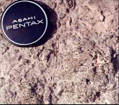
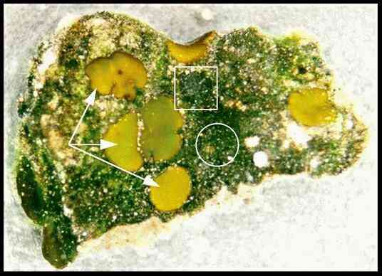
Cryptobiotic crusts are complex communities with many interrelationships that are only partly explored. As an example, we can consider a small part of the crust from the Chihuahuan Desert of Texas.
This crust is completely dry for most of the year, but when a fragment (marked by the white rectangle) was immersed in water the microbial community was reactivated and was found to have at least three components, shown in the image below:
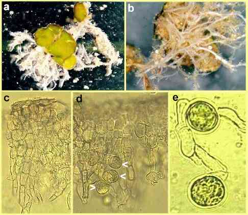
Peltula is a rather unusual lichen consisting of separate lobes, termed squamules, that are connected to one another by a system of fungal hyphae. In (a) and (b) below, we see that these hyphae are aggregated into rope-like structures that lichenologists term "rhizines" but mycologists call them mycelial cords. Their function is to transport nutrients. The mycelial cords were immersed in the carpet of cyanobacteria and penetrated into the underlying sand layers, perhaps obtaining nitrogen and other nutrients from the cyanobacteria. The cords have been dissected away from the sand particles in Figs a (top view) and b (underside). The green lobes of Peltula have a typical lichen structure. The top surface consists of a densely packed fungal tissue (the upper cortex), below which the hyphae have a more normal appearance (c). Cells of a green alga (probably Trebouxia) are found below the lichen surface (arrowheads, d) and are intimately associated with fungal hyphae (e) which gain carbohydrates from the algal cells.
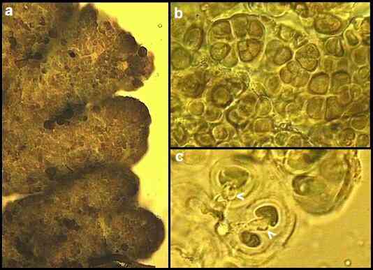
2. The dark-coloured lichen (unidentified) This circular lichen has a lobed margin, seen in (a) below. There is no fungal cortex like that in Peltula. Instead, the upper surface of the lichen consists of clusters of dividing photosynthetic cells embedded in gelatinous sheaths (b). They appear to be cyanobacteria. A network of fungal hyphae extended into the sand from the base of the lichen. These hyphae branched repeatedly near the base of the lichen and the individual short branches (arrowheads in c, below) penetrated into the sheaths surrounding the cyanobacterial cells.
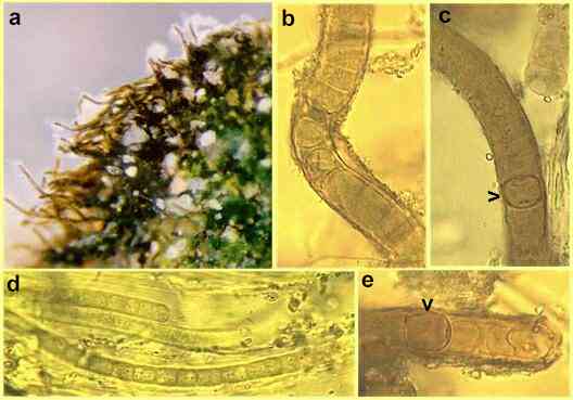
3. The cyanobacteria The predominant cyanobacterium (possibly a species of Scytonema) is seen as a "fringe" of dark-coloured filaments in (a) below and at higher magnification in b, c and e. The filaments are encased in a sheath encrusted with mineral particles (b, e) and contain heterocysts (arrowheads in c and e). Narrower cyanobacterial filaments (d, below) grew from the greener regions of the crust when it was immersed in water for several days.
Other pioneer microbial activities Several bacteria are involved in metal ion transformations in natural environments - they oxidise or reduce sulphur, manganese, iron, etc. to serve in energy-generating processes. These oxidation-reduction activities, whether microbially induced or merely chemical, can be very conspicuous in desert and other dryland regions, where they lead to rock coloration. A classic example is "desert varnish" (not on this server) which recently was shown to be at least partly caused by bacteria (see Magnetite-producing bacteria; not on this server). |
