..
CHAPTER 4: FUNGAL GROWTH This chapter covers the following major topics:
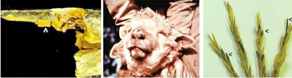
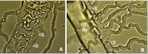
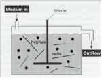
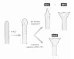
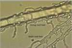
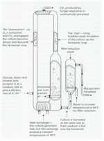
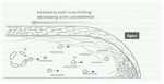
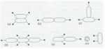
SAMPLE TEXT: In this chapter we focus on the mechanisms of fungal growth, with special reference to the organisation of growth and wall synthesis at the hyphal tip, which is central to understanding the biology of fungi. We also discuss the ways in which hyphal branches arise and orientate themselves for maximum efficiency of nutrient capture. And, we consider the kinetics of fungal growth in batch culture and continuous culture systems, relating this to industrial-scale processes, including the commercial production of Quorn™ mycoprotein – a highly successful fermentation system for producing ‘single-cell protein’. APICAL GROWTH OF FUNGAL HYPHAE Apical growth is the hallmark of fungi. Apart from the fungus-like Oomycota, which have adopted apical growth by a remarkable degree of convergent evolution, no other organisms grow continuously as a tube that extends at the extreme tip by the localised synthesis of wall components. And, arguably, no other group shows such extreme plasticity. The hyphal apex can swell into a balloon-like structure such as a spore or yeast cell, or it can taper to such a degree that it can penetrate a layer of inert gold film or the wall of a host plant by exerting turgor pressure alone. In other circumstances, the fungal hypha can give rise to complex tissues and infection structures, discussed in Chapter 5.
|
|||||||||||||||||||||||