..
MORE IMAGES FROM CHAPTER 3
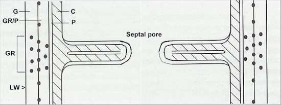
Fig.
3.10. Diagrammatic representation of a simple septum
of Neurospora crassa. The septum develops as an
ingrowing ring from the lateral wall of the hypha.
Associated with this ia a modification of the lateral
wall (LW), including a proliferation of the
glycoprotein reticulum (GR). G = glucan
layer; GR/P = glycoprotein reticulum embedded in
protein; P = protein; C = chitin. [Based on
Trinci, A.P.J. (1978) Science Progress 65,
75-99.]
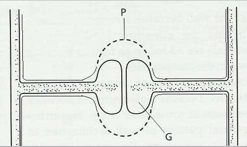
Fig. 3.11. The dolipore septum, found in many members of the Basidiomycota. Large deposits of glucan (G) line the narrow central pore, and specialised perforated membranes termed parenthosomes (P) prevent major organelles from passing through the septal pore. [© Jim Deacon]
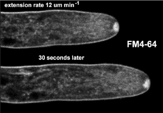
Fig. 3.15a. Confocal images of hyphal tips of Neurospora crassa treated with the steryl dye FM4-64, to detect putative endocytosis. This steryl dye becomes internalised, and strongly labels the Spitzenkörper and the plasma membrane of the hypha. The image shows the same hypha at 30 second intervals, demonstrating that the hypha continued to grow during confocal laser imaging. [© S. Fisher-Parton, R.M.Parton, P.J. Hickey, J. Dijksterhuis, H.A. Atkinson & N.D. Read. Please cite this image as Courtesy of Fisher-Parton et al., The University of Edinburgh]
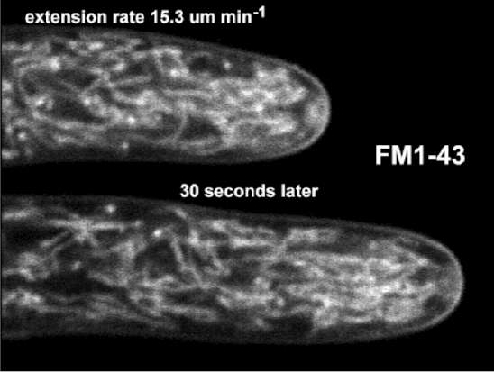
Fig. 3.15b. Confocal images of hyphal tips of Neurospora crassa treated with the steryl dye FM1-43, to detect putative endocytosis. This steryl dye becomes internalised, and strongly labels the mitochondria but only weakly labels the Spitzenkörper. The image shows the same hypha at 30 second intervals, demonstrating that the hypha continued to grow during confocal laser imaging. [© S. Fisher-Parton, R.M.Parton, P.J. Hickey, J. Dijksterhuis, H.A. Atkinson & N.D. Read. Please cite this image as Courtesy of Fisher-Parton et al., The University of Edinburgh]
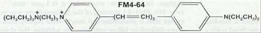
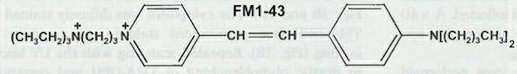
Fig. 3.15c. Formulae of the two steryl dyes, FM4-64 and FM1-43. The images shown in Figs 3.15a and 3.15b clearly show the power of modern techniques for investigating the behaviour of living fungal hyphae.
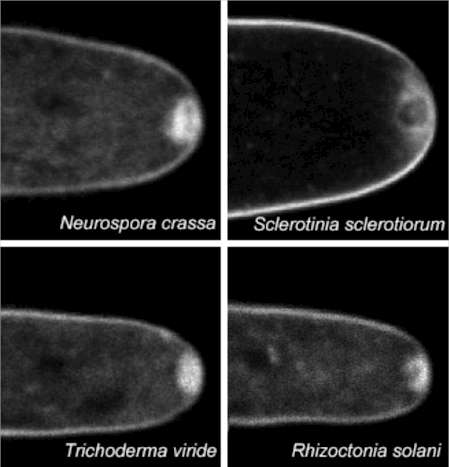
Fig. 3.16. Dye labelling (FM4-64) of the cytoplasmic membrane and Spitzenkörper of different fungi, including Ascomycota (Neurospora crassa), Basidiomycota (Sclerotinia and Rhizoctonia), and mitosporic fungi (Trichoderma viride). Phycomyces (Zygomycota) also showed similar labelling (not shown in this image). [© S. Fisher-Parton, R.M.Parton, P.J. Hickey, J. Dijksterhuis, H.A. Atkinson & N.D. Read. Please cite this image as Courtesy of Fisher-Parton et al., The University of Edinburgh]
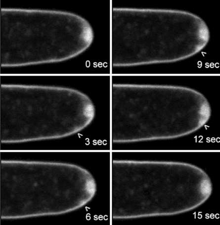
Fig. 3.17: Confocal images of an FM4-64-stained satellite Spitzenkörper (arrowhead) of Botrytis cinerea showing a time course (seconds) of different stages in its formation, migration and fusion with the main Spitzenkörper. [© S. Fisher-Parton, R.M.Parton, P.J. Hickey, J. Dijksterhuis, H.A. Atkinson & N.D. Read. Please cite this image as Courtesy of Fisher-Parton et al., The University of Edinburgh]
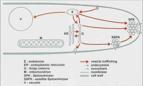
Fig.
3.18. Hypothetical model of the organisation of the
vesicle-trafficking network in a growing hypha, based on
the pattern of FM4-64 staining.
|
|||||||||||||