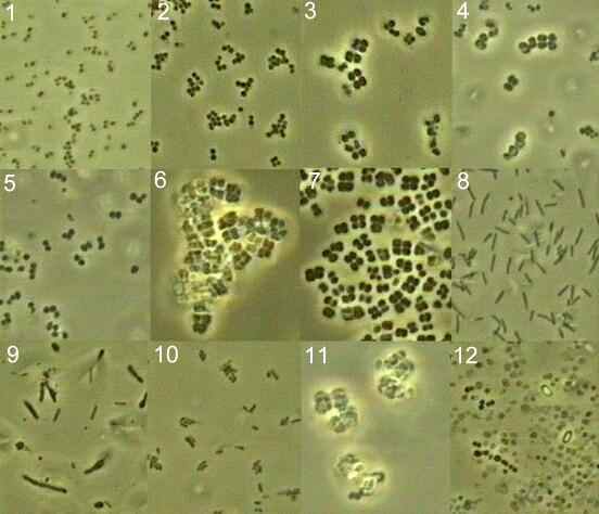
Colony 1
Small cocci occurring
singly or in small groups.
Return to bacterial shapes
Colony 2

Cocci in small chains
or clusters.
Return to bacterial shapes
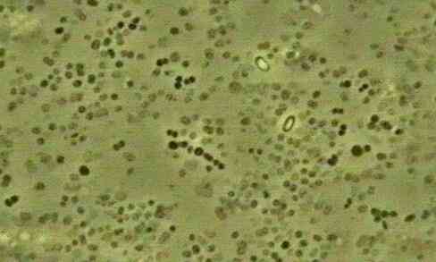
Colony 3

Large cocci, forming
tetrads.
Return to bacterial shapes
Colony 4

Large cocci, forming
tetrads.
Return to bacterial shapes
Colony 5

Cocci occurring singly
or in small clusters.
Return to bacterial shapes
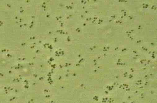
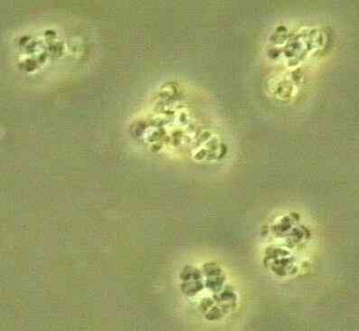
Colony 6
Large cocci with
three-dimensional planes of cell division, giving regular
'packets' of cells, typical of Staphylococcus.
Return to bacterial shapes
Colony 7

Large cocci, forming
tetrads.
Return to bacterial shapes
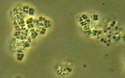
Colony 8
Long, thin rods
Return to bacterial shapes
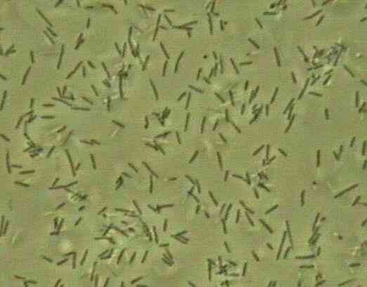
Colony 9
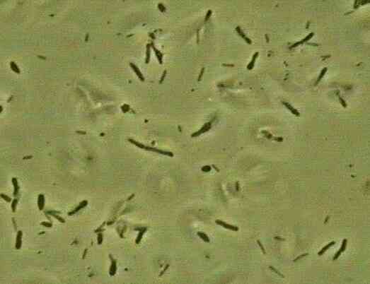
Large pleomorphic
(variably shaped) rods
Return to bacterial shapes
Colony 10
Short rods
Return to bacterial shapes
Colony 11
Large cocci with
three-dimensional planes of cell division, giving
'packets' of cells, typical of Staphylococcus.
Return to bacterial shapes
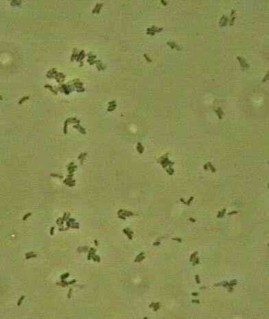
Colony 12
Mixed population of
bacteria enveloped in slime (which accounts for the poor
phase-contrast), including Streptococcus (chains
of cocci) and two phase-bright spores.
Return to bacterial shapes
|