..
MORE IMAGES FROM CHAPTER 9:
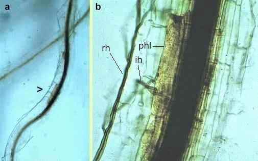
Fig 9.11 A wheat root at different magnifications, infected by the take-all fungus, Gaeumannomyces graminis. The fungus grows on the root surface as dark ‘runner hyphae’ (rh) then invades the root cortex by infection hyphae (ih), destroying the phloem (phl) and causing intense discoloration and blockage of the xylem. [© Jim Deacon]
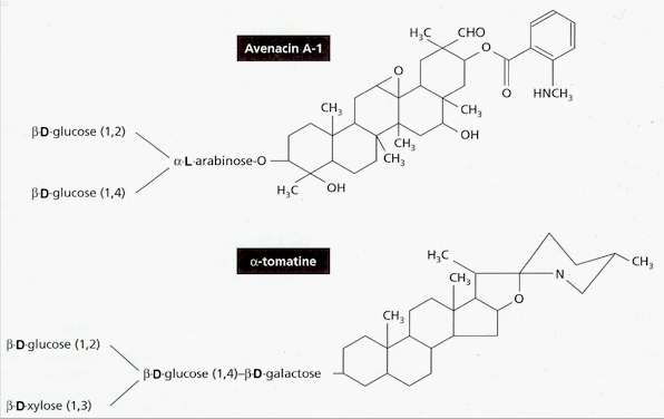
Fig 9.12 Structures of two saponins that are pre-formed resistance compounds in plants: avenacin in roots of oats, and a-tomatine in tomato. Pathogens with the appropriate enzymes can detoxify these compounds by cleaving some of the terminal sugar residues. [© Jim Deacon]
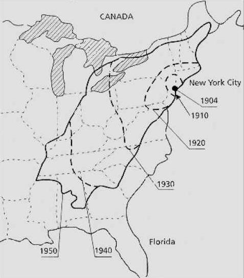
Fig 9.13 Recorded spread of chestnut blight caused by Cryphonectria parasitica after it was first recorded in New York in 1904.
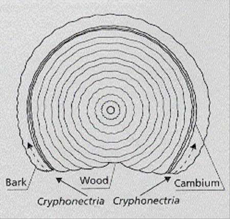
Fig 9.14 Diagram of a cross-section of a trunk with a spreading canker caused by Cryphonectria parasitica. This fungus produces airborne spores that infect through wounds, then progressively girdles the trunk by growing in the cambium. [© Jim Deacon]
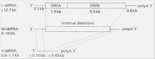
Fig 9.15 Double-stranded RNA of hypovirulent strains of C. parasitica. Large (L) dsRNA is the full-length molecule comprising two conserved end regions and a central coding region of two open reading frames (ORF A and ORF B) which confer hypovirulence. Medium (M) and small (S) dsRNAs are also commonly found in hyphae. They are internally deleted copies of the L-dsRNA.
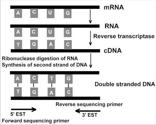
Fig 9.16 Procedure for generating cDNA from messenger RNA, then producing expressed sequence tags (ESTs) from either the 5’ or 3’ end of cDNA.
|
||||||||||||