..
IMAGES FROM CHAPTER 8: ENVIRONMENTAL CONDITIONS FOR GROWTH, AND TOLERANCE OF EXTREMES
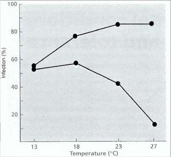
Fig 8.1 Effect of temperature on infection of wheat seedlings by the take-all fungus of cereals, Gaeumannomyces graminis, in sterilised soil (top line) and unsterile soil (bottom line). [From Henry, 1932] [© Jim Deacon]
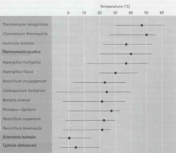
Fig 8.2 Approximate temperature ranges and optima (o) for growth of some representative fungi [© Jim Deacon]
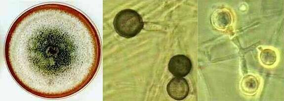
Fig 8.3 Thermomyces lanuginosus, a mitosporic thermophilic fungus. Left: 10-day-old colony on malt-extract agar. Centre and Right: large conidia which are darkly pigmented at maturity and are borne singly on short hyphal branches. [© Jim Deacon]
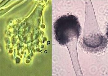
Fig 8.4 Aspergillus fumigatus, a thermotolerant mitosporic fungus. Left and centre: Flask-shaped phialides (p) produce conidia (c) on the inflated, club-shaped heads of conidiophores. Right: Clinical specimen, stained to show the hyphae of A. fumigatus in an aspergilloma of the lungs. [© Jim Deacon]
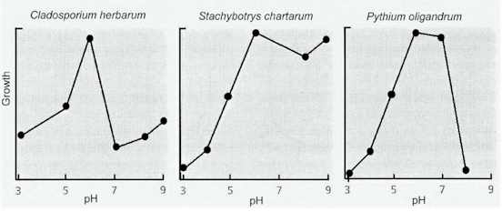
Fig 8.5 The pH growth response curves of three representative fungi in laboratory culture [© Jim Deacon]
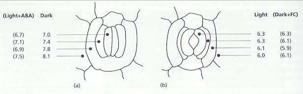
Fig 8.6 pH of the leaf surface on individual cells around the stomata of Commelina communis, measured with a pH electrode.(A) pH measured when the stomata were closed - either by incubating the leaves in darkness or (in brackets) by treatment with abscisic acid in the light. (B) When stomata were open - either by incubation in the light or (in brackets) by treatment with fusicoccin in darkness. Based on data in Edwards MC & Bowling DJF (1986). The growth of rust gern tubes towards stomata in relation to pH gradients. Physiological & Molecular Plant Pathology 29, pp 185-196.
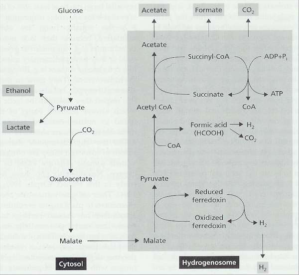
Fig 8.8 Diagrammatic representation of the mixed-acid fermentation of the rumen chytrid Neocallimastix. The end products of this fermentation are shown in the small shaded boxes. Part of the fermentation occurs in the cytosol, part in the hydrogenosome. Some of the details are known (Orpin, 1993; Marvin-Sikkema et al., 1994); others are assumed, based on knowledge of the mixed-acid fermentation of some enteric bacteria. [Based on A.P.J.Trinci, D.R.Davies, K.Gull et al.(1994), Mycological Research 98, 129-152]
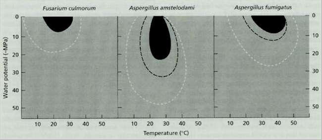
Fig 8.10 Growth rate isopleths for three fungi that cause spoilage of cereal grains. Data for Aspergillus spp. from G.Ayerst (1969), Journal of Stored Products Research 5, 127-141, and for F. culmorum from N.Magan & J.Lacey (1984) Transactions of the British Mycological Society 82, 71-81.
Fig 8.11 Combinations of temperature and water potential that support aflatoxin production by Aspergillus flavus, ochratoxin A production by Penicillium verrucosum and patulin production by Penicillium expansum. Based on data in MD Northolt & LB Bullerman (1982). Prevention of mould growth and toxin production through control of environmental conditions. Journal of Food Production 45, pp 519-526.
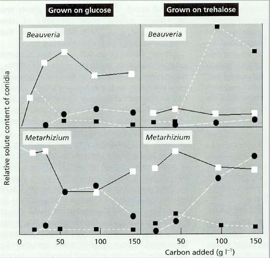
Fig 8.12 Changes in the compatible solutes of the conidia of two insect-pathogenic fungi (Beauveria bassiana and Metarhizium anisopliae). Conidia were harvested from agar plates that contained increasing levels of glucose or trehalose. The compatible solutes found in the conidia were: mannitol (white squares), erythritol or arabitol (black circles), and trehalose (black squares) [Adapted from Hallsworth & Magan (1994) Microbiology 140, 2705-2713].
Fig 8.13 Examples of darkly pigmented spores of fungi that commonly grow on senescing leaves and stems of plants, and that also grow in condensation on bathroom and kitchen walls. These fungi are often termed dematiaceous hyphomycetes (meaning that they have darkly pigmented hyphae and spores). The left-hand panel shows several spores of Alternaria (30-40 micrometres diameter) viewed by phase-contrast or bright-field microscopy. The right-hand panel shows spores of Cladosporium spp. and related fungi, which are smaller (about 10-15 micrometres diameter) but are shown at a different scale.[© Jim Deacon] |
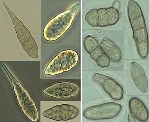
|||||||||||||