..
MORE IMAGES FROM CHAPTER 5
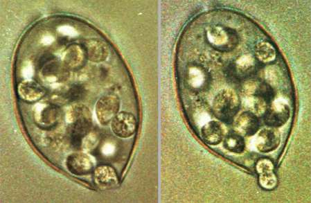
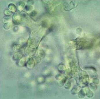
Fig. 5.17. Detached sporangia of Phytophthora infestans, incubated at 12oC. The sporangial contents cleave to produce zoospores, then the apical papillum of the sporangium breaks down and the zoospores are released by squeezing through the narrow opening . [© D. Grayson, A. Hill & Jim Deacon, The University of Edinburgh]
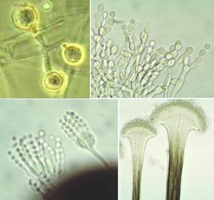
Fig. 5.18. Some examples of conidial development. Top left: single conidia of Thermomyces lanuginosus, produced at the ends of short hyphal branches. Top right: branched chains of conidia of Monilinia (the asexual stage of Sclerotinia fructigena) produced by a repeating budding-like process before the spores separate by the development of septa. Bottom left: Penicillium expansum conidiophores with phialides that produce chains of spores. Bottom right: coremia of Ophiostoma ulmi; the conidiophores are aggregated to form stalk-like structures that produce masses of spores at their tips (but most of the spores have been dislodged during preparation of this specimen. [© Jim Deacon]
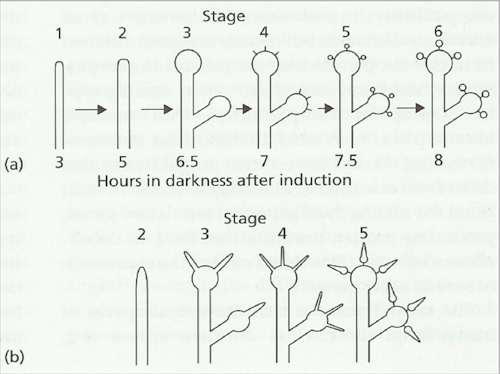
Fig. 5.19. Diagrammatic representation of stages in the development of conidia of Botrytis cinerea. (a) After exposure to near-ultraviolet (NUV) irradiation the aerial hyphae are transformed into branched conidiophores which swell at the tips to form small globose vesicles. Conidia are formed on minute projections from these vesicles, so the mature clusters resemble bunches of grapes – see Fig. 5.20. (b) A short exposure to blue light at different times after triggering of development by NUV causes development to be arrested. The fungus then switches back to a hyphal growth form – for example, the swelling of the immature conidia stops and the fungus produces narrow hyphal outgrowths from the immature conidia. [Based on Suzuki et al., 1977]
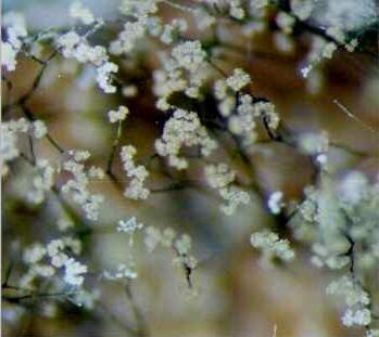
Fig. 5.20 Botrytis cinerea, which commonly causes ‘grey mould’ of soft fruits. Left: low-power view of the characteristic branching conidiophores which bear clusters of grey conidia at their tips. Right: close-up showing the bulbous tips of the conidiophore branches, bearing immature conidia.
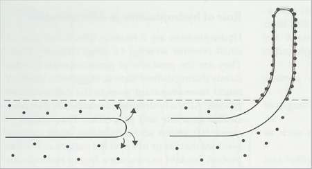
Fig. 5.21. Release of hydrophobin proteins from hyphae of Schizophyllum commune submerged in a culture medium (left), and spontaneous polymerisation of hydrophobins into water-repellent films (right) when the hyphae emerge into an aerial environment. [Based on a diagram in Wessels, 1996]
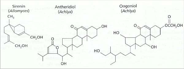
Fig. 5.22. Three pheromones that regulate sexual reproduction in zoosporic organisms. Sirenin is released by female gametes of Allomyces spp. Antheridiol and oogoniols regulate sexual attraction and mating in Achlya spp. (Oomycota) and possibly in other Oomycota.
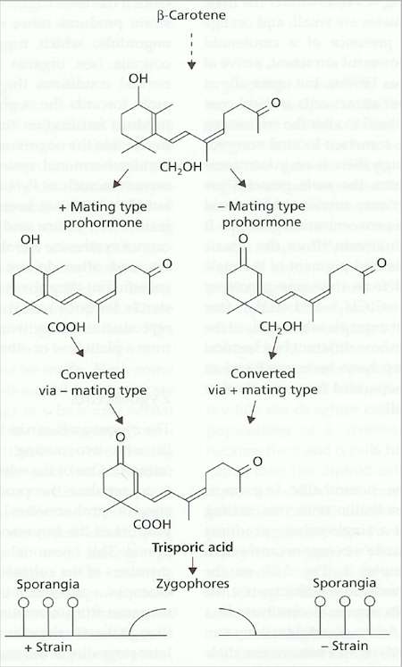
Fig. 5.23. The trisporic acid hormonal system of Zygomycota. See text for details. |
||||||||||||