MORE IMAGES FROM CHAPTER
10:
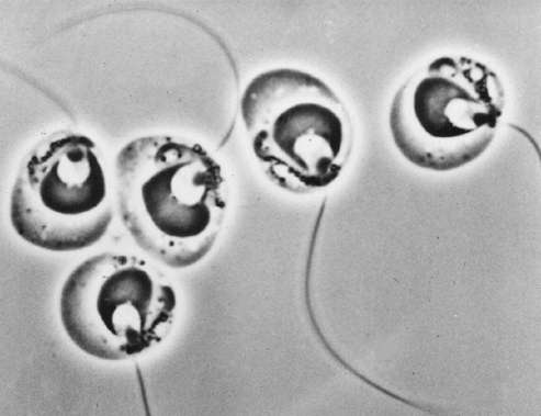
Fig 10.12a Zoospores of Blastocladiella emersonii (Chytridiomycota) each with a smooth, posterior whiplash flagellum (n = nucleus; nc = nuclear cap; m = mitochondrion; flag = flagellum) [Image supplied by Courtesy of M. S. Fuller].
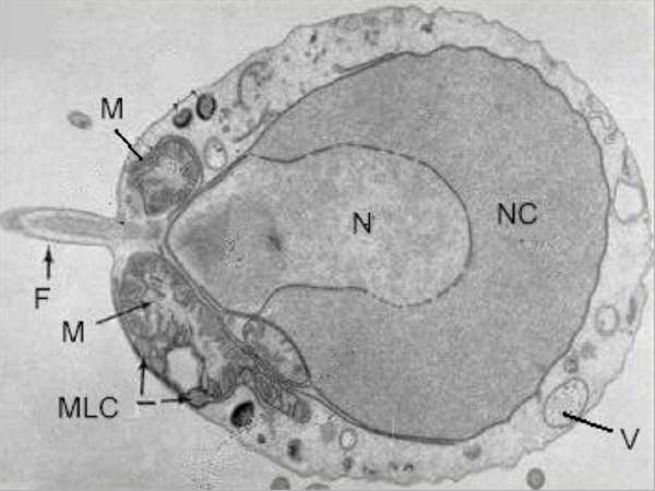
Fig 10.12b: Electron micrograph of a longitudinal section of the zoospore of B. emersonii. The zoospore plasma membrane is continuous with the flagellar membrane (F) but only part of the flagellum is seen in this section. V = vacuole; mlc = microbody-lipid globule complex. [Image supplied by courtesy of M. S. Fuller; from Reichle & Fuller, 1987]
Fig 10.13 Top: scanning electron micrograph of a zoospore of Phytophthora (Oomycota) with a posterior whiplash flagellum and a shorter, anterior tinsel-type flagellum. [Image courtesy of M. S. Fuller] Bottom: diagrammatic representation of the zoospore, showing the insertion of the flagella in the ventral groove (shaded) and the location of the nucleus (N) and water-expulsion vacuole (W) [© Jim Deacon]
Fig 10.15 Close-up of a part of the zoospore surface of Pythium aphanidermatum, showing the laminate peripheral cisternae that will subsequently become vesicles for release of the cyst wall. [© Jim Deacon]
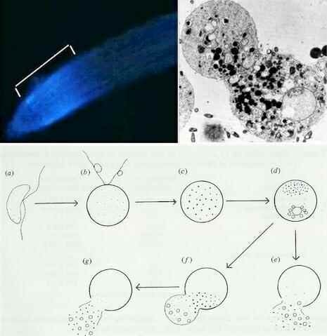
Fig 10.17 Top left: Oat root tips with natural blue autofluorescence caused by the presence of avenacin. Top right: a Pythium zoospore undergoing disorganisation and lysis in the presence of avenacin. Lower sequence: Responses of wall-less zoospores of Oomycota to saponins such as avenacin or ß-aescin. (a) motile zoospore; (b) immobilisation and rounding-up; (c) development of phase-dark granules, (d) localisation of granules and development of vacuoles; (e) lysis; (f,g) ballooning followed by lysis. Disruption of the zoospores usually occurs within 5-10 minutes. [From Deacon & Mitchell, 1985] [© Jim Deacon]
Fig 10.18 Zoospore tracks of Oomycota, captured as negatives on photographic film during a 5-second exposure and showing frequent random turns in the absence of an attractant. [© Jim Deacon]
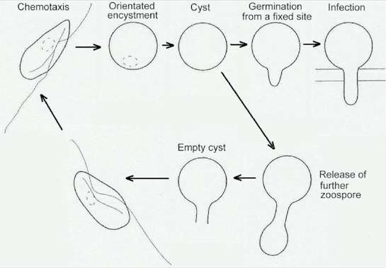
Fig 10.19 The homing and docking sequence of zoospores. See Chapter 10 for details. [© Jim Deacon]
Fig 10.20 Accumulation and encystment of Pythium zoospores on wheat roots. Left: zoospores (z) accumulating in large numbers on the surface of a ball of root tip mucilage; some root cap cells are indicated (rc). Right: zoospores (z) accumulating and encysting in the elongation zone of wheat roots. In this case the root had been coated with a double layer of calcium alginate gel to block any specific receptor compounds on the root surface. [© Jim Deacon]
Fig 10.21a A suspension of zoospores of Phytophthora parasitica was incubated in the presence of a fluorescent probe (fura-2) that measures Ca2+ concentration in the suspending fluid. The zoospore suspension was then vortexed (70 second interruption) to induce zoospore encystment and the external Ca2+ measurements were resumed. The trace shows that zoospore encystment caused an immediate drop in external Ca2+ (signifying Ca2+ uptake by zoospores), then a progressive Ca2+ release from the cysts, which germinated within 90 min. [© Jim Deacon]
Fig. 10.21b,c. In identical experiments, the addition of lanthanum or verapamil (both of which are Ca2+ channel blockers) prevented the release or uptake of Ca2+. The vortexed cells were immobilised but did not produce cyst walls. Another calcium-modulator, TMB-8, caused the cells initially to behave like the controls (an early uptake of Ca2+ after vortex-treatment) but with no subsequent release of Ca2+ and no germination. TMB-8 is known to block the release of Ca2+ from intracellular stores. [All data from Warburton & Deacon, 1998; [© A. Warburton & Jim Deacon] |
|||||||||||||