..
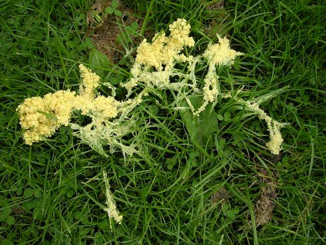
FOCUS TOPIC: PLASMODIAL AND CELLULAR SLIME MOLDS SLIME MOLDS ARE NOT FUNGI. Instead, they are classified within the "Kingdom" of protists (Protoctista) that includes all the small organisms that posess nuclei - the amoebae, protozoa, algae, slime moulds, etc. Molecular phylogeny based on the comparison of genes that encode the small subunit ribosomal RNA (see Chapter 1) can be used to place all these organisms within a phylogenetic framework. But this process has only just begun and so, for the time being, all these small nucleate organisms are grouped within the Protoctista. Even the slime molds are not a natural phylogenetic grouping, but they have a number of features in common, including the fact that at some stage in their life cycle they produce naked, amoeba-like stages which engulf food particles such as bacteria or other small organic materials. As the amoeba-like stages grow they undergo nuclear division, which can lead eventually to very large multinucleate masses of naked (wall-less) protoplasm. Depending on environmental conditions, these multinucleate masses can develop into large resting structures (sclerotia) or they can differentiate into spore-bearing (fruiting) structures. The life cycle is completed when the spores are dispersed, usually by wind, and then germinate to release amoeboid cells. Three main groups of slime molds are currently recognised: (1) the plasmodial slime moulds, termed the Myxomycota (or members of the Class Myxomycetes), which can grow to macroscopic proportions, consisting of a huge mass of protoplasm containing many millions of nuclei; (2) the cellular slime moulds such as Dictyostelium (dictyostelids - sometimes called the Dictyosteliomycota; see Chapter 2); (3) the acrasid slime molds (Acrasiomycota) which resemble the dictyostelids but are more variable and may not be closely related to one another. Here we will focus initially on the plasmodial slime molds - the Myxomycota - of which there are more than 500 species. These organisms remain hidden for most of the year, growing in moist soil, on decaying wood or in dung. But in response to environmental triggers such as exhaustion of the food base or exposure to light, they can migrate to the surface and produce conspicuous spore-bearing structures. A classic example is the many-headed slime-mold, Muciturbo crustacea (Fig. 1).
Fig. 1. The plasmodial slime mold, Muciturbo crustacea, which emerged from a naked protoplasmic mass below ground and migrated to the surface of the author's lawn, where it began to differentiate into its sporing stage. This plasmodium was about 15 cm diameter. [© Jim Deacon]
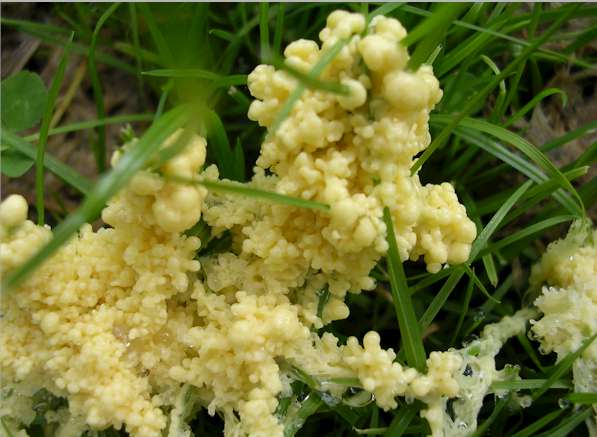
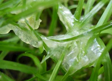
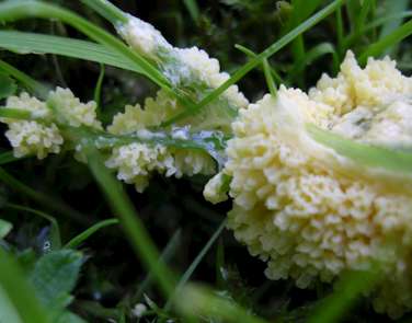
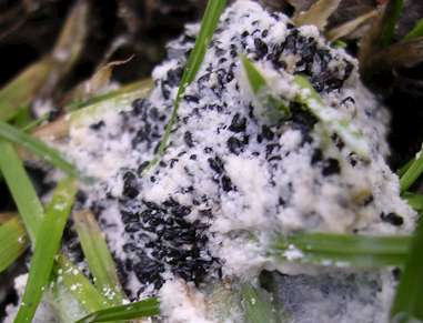
Fig. 2. Close-up of part of the plasmodial slime mold, M. crustacea. Note the undifferentiated 'slimy' protoplasm in the lower right corner of the image. [© Jim Deacon] Muciturbo crustacea, like many plasmodial slime molds, produces small clusters of spore-bearing heads at the top of short stalks which are composed of dead amoebal cells. But some other plasmodial slime molds produce larger spore-bearing structures termed aethalia (singular, aethalium). Figure 3 (below) shows three stages in the development of M. crustacea, emerging from soil onto some blades of grass. Each image is about 5 cm diameter. The sequence of images was taken over a 5-day period. The initial jelly-like plasmodium migrated from the soil onto the grass leaves, and them developed into a rosette-like structure. After several days this transformed into a white amorphous mass with several black spore-bearing structures that eventually release spores. [© Jim Deacon]
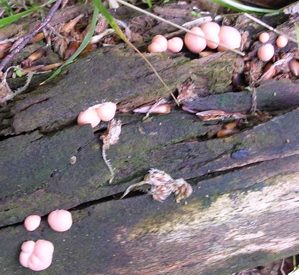
Fig. 4.(below) Immature, pink aethalia of the slime mold, Lycogala epidendrum, growing on a the wood of a fallen, decaying pine tree. Each fruitbody is about 1 - 1.5 cm diameter. At this immature stage, the fruitbodies have a soft, spongy consistency and consist of a large protoplasmic mass. [© Jim Deacon]
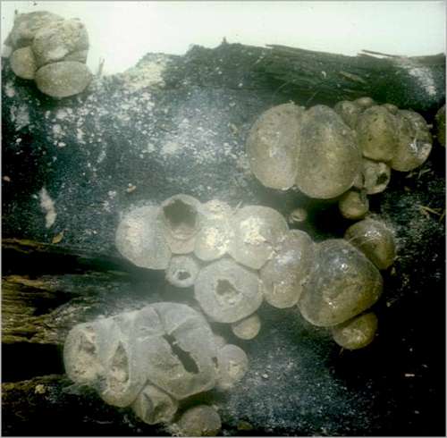
Fig. 5. Mature aethalia of the slime mold, Lycogala epidendrum, about 3-5 days after the image in Fig. 3 was taken. The aethalia have now differentiated and consist of a thin, gray, papery sac containing many walled spores. These spores are released when the fruitbody develops an apical split. Each fruitbody is about 1 - 1.5 cm diameter. [© Jim Deacon]
|