..
MORE IMAGES FROM CHAPTER 2
Fig.
2.31. The ‘ink caps’ (Coprinus spp.)
and related fungi, commonly found on animal dung or in
composts. Typically, the basidiocarps develop rapidly but
last for only a short time. The cap containing the gills
then senesces (deliquesces) from the base upwards,
dripping an inky fluid containing the black
basidiospores. Mycological enthusiasts have been known to
write with this material, but that was before computers! Left:
Coprinus comatus (known as shaggy cap, or
lawyer’s wig), about 15 cm tall at maturity. Centre:
Similar basidiocarps 2-3 days after maturity and showing
deliquescence. Right: a related genus, Psathyrella,
growing on an agar plate. Coprinus and related
species are among the few mushroom-producing fungi that
can be grown easily in agar culture. They have been used
extensively in genetical and developmental studies.
Fig. 2.32 The unmistakable 'fly agaric', Amanita muscaria, with a cap about 12 cm diameter. Note the swollen, bulb-shaped volva at the base of the stalk, and the ring of shredded tissue just below the cap. The cap itself bears white scales - the remnants of the membrane that initially enclosed the whole cap in its immature stage. A. muscaria is extremely poisonous. [© Jim Deacon]
Fig. 2.33. Phallus impudicus, the stinkhorn, about 12 cm long. This fungus often develops from rotting wood below ground. It first appears as a gelatinous ‘egg’, about the size of a golf ball (see the lower part of the left-hand image) but then elongates as a spongy stipe with a distinctive cap. The cap is covered initially with a putrid-smelling, dark olive slime containing the spores. Flies are strongly attracted by the smell, land on the cap, and carry the slime away as it sticks to their legs, serving to disperse the spores. Even when the slime and spores have gone, flies and other insects are still strongly attracted by the odour from this fungus (see the right-hand image). [© Jim Deacon]
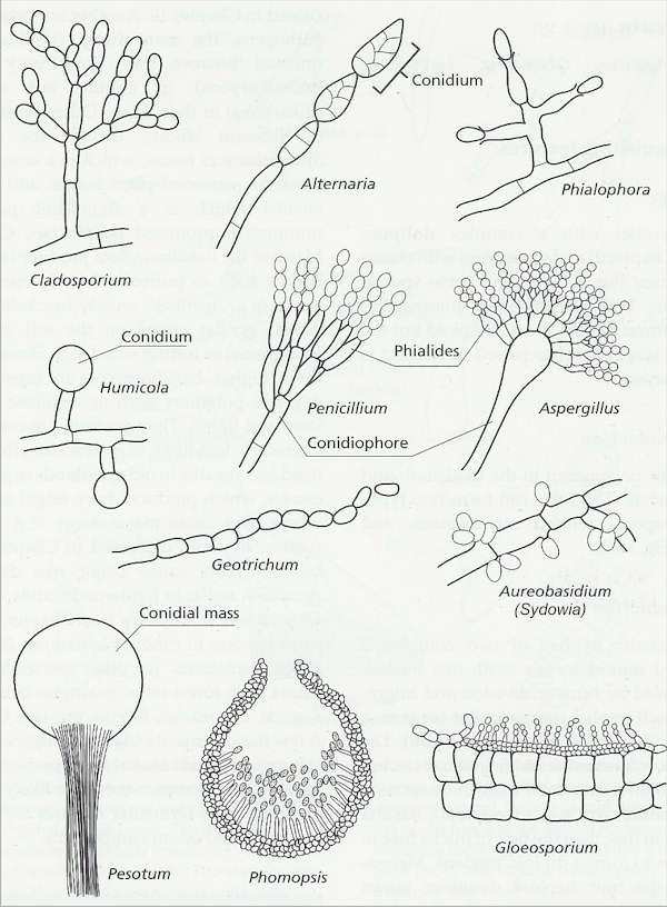
Fig. 2.34. Some representative methods of conidium production in mitosporic fungi. [© Jim Deacon] |
|||||||||||


