..
MORE IMAGES FROM CHAPTER 2
Fig. 2.13. Left: A developing perithecium of Sordaria macrospora which, at maturity, will have a pore at its tip to release the ascospores. Right: several asci in different stages of development within the perithecium; each ascus contains 8 ascospores, about 20 µm diameter. [© N.D. Read & K.M.Lord (1991), The University of Edinburgh] From: Experimental Mycology 15, 132-139.
Fig. 2.14. Diagrammatic representation of some fruiting structures of Ascomycota. (a) A single ascus containing 8 ascospores which, at maturity, will be shot forcibly through the narrow apical pore. (b) A cup-shaped apothecium containing many asci interspersed with sterile ‘packing’ hyphae (paraphyses). The paraphyses help to laterally constrain the developing asci so that they project beyond the surface of the cup to release their spores. (c) A cleistothecium (closed ascocarp) such as that of Eurotium, the sexual stage of some Aspergillus spp. The asci, each with 8 ascospores, are released when the cleistothecium wall breaks down at maturity to release the ascospores. [© Jim Deacon]
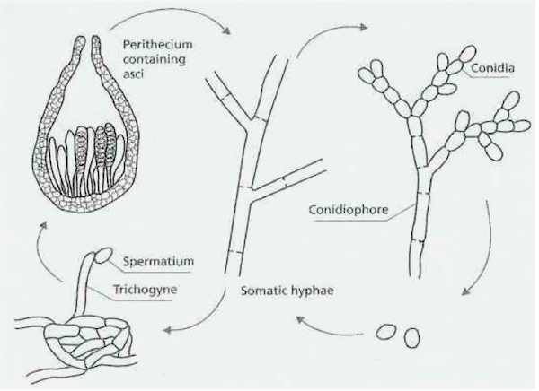
Fig. 2.15. Stages in the life cycle of Neurospora crassa and many similar fungi in the Ascomycota. Like many members of the Ascomycota, N. crassa, has a sexual stage, leading to the production of asci containing ascospores. N. crassa is heterothallic, requiring strains of different mating types (termed A and a) for sexual development. The female sex organ, termed an ascogonium, is often a coiled, multinucleate hypha with a receptive trichogyne. It is fertilised either by contact with a spermatium (a small uninucleate cell that cannot germinate) or by a conidium. Then the ascogonium produces an ascogenous hypha that will eventually give rise to the asci. The process by which this happens is shown in Fig. 2.16. [© Jim Deacon]
Fig. 2.16. Early stages in the production of asci from an ascogenous hypha. (a) The ascogenous hypha grows for some distance then curls back at its tip to form a crosier. (b) Synchronous nuclear division occurs as the crosier bends back upon itself. (c) Septa are laid down so that the penultimate cell (located at the top) has one nucleus of each mating type. This cell will extend and will give rise to the first ascus, when the nuclei fuse and then undergo meiosis and one round of mitosis, to produce 8 ascospores. Meanwhile, the tip cell of the crosier fuses with the ascogenous hypha, and the nucleus is transferred. (d) A new branch develops from the fused cell, and this branch forms a further crosier, to repeat the whole process. Thus, eventually, a large number of ascus initials are produced, which will give rise to a large number of asci. The final stage in this process is the development of a fruitbody tissue surrounding the asci, as shown in Fig. 2.17. The haploid ascospores are either A or a mating type, and eventually germinate to produce haploid colonies, to complete the life cycle. [© Jim Deacon]
Fig. 2.17. Taphrina populina, a plant-parasitic member of the Archaeascomycota which causes yellow blister of poplar leaves (Populus spp.). The fungus grows as a mycelium in the plant leaves, causing leaf distortion, and it produces naked asci which project from the surface of the leaves. Within the asci the ascospores often bud, so that the asci become filled with a mixture of ascospores and budding yeast cells (particularly evident in the top ascus). Click HERE for the leaf symptoms of Taphrina populina. [© Jim Deacon]
Fig. 2.18. Generalised life cycle of Basidiomycota, exemplified by Agaricus [© Dr Maria Chamberlain, The University of Edinburgh]
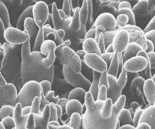
Fig. 2.19. Scanning electron micrograph of the basidia of a mushroom, Coprinus cinereus. Note the inflated basidia, each with four sterigmata (stalks), and the basidiospores, some of which have already been released, or collapsed during preparation of the specimen. [© Dr Chris Jeffree, The University of Edinburgh]
Fig. 2.20 The role of clamp connections in maintaining a regular dikaryon in members of the Basidiomycota.[© Jim Deacon] |
|||||||||||


