..
CHAPTER 16: FUNGAL PATHOGENS OF HUMANS
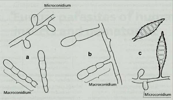
Fig. 16.1. Spores of the three common genera of dermatophytic fungi. (a) Macroconidia (about 50 µm long) and microconidia (about 4 µm) of Trichophyton spp. (b) Macroconidia of Epidermophyton spp, which do not produce microconidia. (c) Spindle-shaped macroconidia, and microconidia of Microsporum spp. [© Jim Deacon]
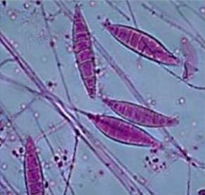
Fig. 16.2a: Spindle-shaped macroconidia, and microconidia, of Microsporum spp. [Reproduced by courtesy of the Canadian National Centre for Mycology; http://www2.provlab.ab.ca/bugs/webbug/mycology/dermhome.htm]
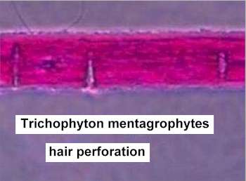
Fig16.2b: Enzyme-mediated penetration of a hair by hyphae of Trichophyton mentagrophytes. [Reproduced by courtesy of the Canadian National Centre for Mycology; http://www2.provlab.ab.ca/bugs/webbug/mycology/dermhome.htm]
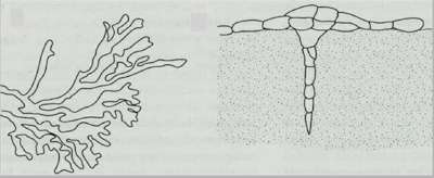
Fig. 16.3. Left: Flattened fronds of hyphae of a dermatophyte, growing in planes of weakness within a stratified substrate such as skin flakes. Right: Diagram of a perforating organ, similar to that in Fig 16.2b.[© Jim Deacon]
Fig. 16.4. Pseudomycelium of Candida albicans in nutrient-limiting conditions. Yeast cells are produced instead of fungal branches at the septa. [© Jim Deacon]
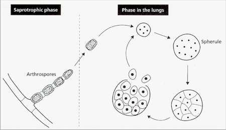
Fig. 16.5. Infection cycle of Coccidioides immitis, which grows in desert soils. In its saprotrophic phase, the fungus produces hyphal branches that undergo multiple septation to form a chain of cells. Then the nutrients are withdrawn from every alternate cell, to supply nutrients for the remaining cells (arthrospores) which develop thick walls. Arthrospores carried in wind-blown soil enter the lungs, germinate and produce a multinucleate spherule. At maturity, the cytoplasm cleaves around the individual nuclei, and the spherule releases uninucleate cells that can develop into further spherules.
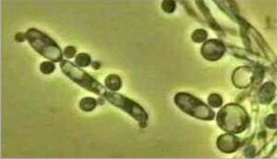
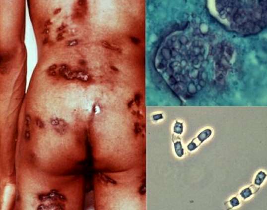
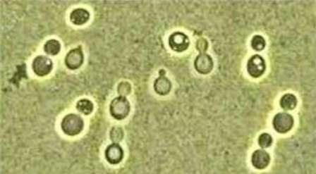
Fig. 16.6. Left: A patient showing the disseminated stage of disease (coccidioidomycosis). Top right: spherules. Bottom right: chains of arthrospores interspersed with empty cellular compartments. [Images reproduced by courtesy of the DoctorFungus website; http://www.doctorfungus.org/]
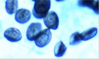
Fig. 16.7. Typical appearance of the budding phase of Paracoccidioides brasiliensis: the large parent yeast-like cell produces multiple buds that remain attached to the mother cell.
Fig. 16.8. Cryptococcus neoformans and its sexual stage, Filobasidiella neoformans. The budding yeast cells (Y-phase) of opposite mating types conjugate and form a dikaryotic cell. This forms a mycelium on which the inflated basidia develop. Meiosis in the basidia leads to the production of four haploid nuclei, which migrate to the tips of the basidia. Then chains of haploid basidiospores are produced, and these germinate to give the haploid Y-phase cells. [© Jim Deacon]
Fig. 16.9. Cells of a Cryptococcus species (C. albidus) viewed by phase-contrast microscopy in the presence of India ink, which clearly shows the rigid polysaccharide capsules. [© Jim Deacon]
Fig. 16.10. Saucer-shaped cysts of Pneumocystis jiroveci [Reproduced with permission from Centers for Disease Control and Prevention, Image library] http://www.dpd.cdc.gov/dpdx/HTML/ImageLibrary/Pneumocystis_il.htm] |
|||||||||||