..
IMAGES FROM CHAPTER 13: FUNGAL SYMBIOSIS
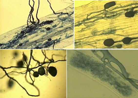
Fig. 13.1. The principal features of arbuscular mycorrhizal (AM) fungi, observed by clearing the root tissues with strong alkali and then staining roots with the fungal dye, trypan blue. Top left: A root heavily colonised by AM fungi, with hyphae that radiate into the soil. Top right: When observed through the depth of the root cortex, AM fungal hyphae are often seen to run parallel to the root axis, growing between the root cortical cells. These hyphae are irregular, with constrictions and bulges, quite unlike the hyphae of most other fungi. They frequently produce large, swollen vesicles within the root tissues. Bottom left: some of the external hyphae and hyphal aggregates produce clusters of spores in the soil. Bottom right: Some of the root cortical cells are penetrated by hyphae that branch repeatedly to produce intricately branched arbuscules, often completely filling the root cells. [© Jim Deacon]
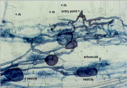
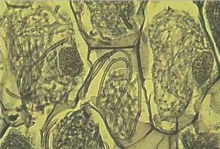
Fig. 13.2. Part of a clover root, partly crushed and cleared of protoplasm by treatment with strong alkali, then stained with trypan blue to show the distribution of fungal structures in the root. The main features shown are root hairs (rh) an entry point with a characteristic diamond-shaped swelling equivalent to an appressorium, large swollen vesicles, and ‘fuzzy’ arbuscules. [© Jim Deacon]
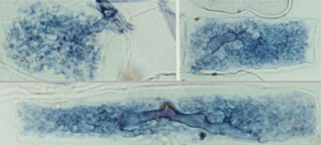
Fig. 13.3. Single arbuscules of AM fungi growing within root cells. The arbuscules enter individual root cells from intercellular hyphae, then branch dichotomously and repeatedly so that most of the cell volume is filled with finely branched AM hyphae, providing a large area of interface for potential nutrient exchange between the fungus and its plant host. [© Jim Deacon]
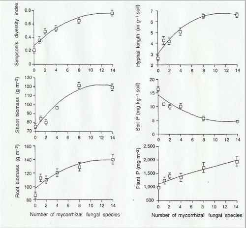
Fig. 13.4. An increase in the number of different arbuscular mycorrhizal fungi in a soil leads to an increase in plant productivity and plant biodiversity. [From van der Heijden et al., 1998, with permission from the publisher]
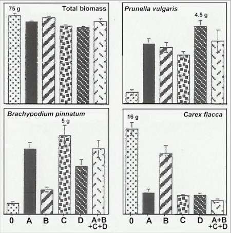
Fig. 13.5. Total biomass and the biomass of three representative plant species grown in soil with no mycorrhizal fungus (0) or with four separate AM fungal species (A,B,C,D) or a combination of all four AM species. Note that the vertical scale of each histogram is different but the largest biomass is shown in each case. [Data from van der Heijden et al., 1998, with permission from the publisher, but only some of the plant species are shown in this Figure]
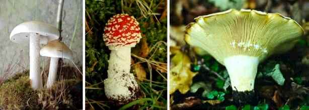
Fig. 13.6. The fruitbodies (basidiocarps) of some representative fungi that form ectomycorrhizas with forest trees. From left to right: Hebeloma sp. growing on the roots of oak; Amanita muscaria growing on birch roots; Lactarius sp. (which, typical of the genus, secretes a milky fluid when the gills are damaged) growing on birch roots. [© Jim Deacon]
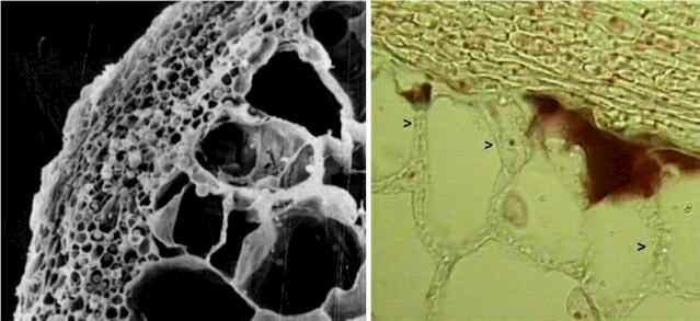
Fig. 13.7. Left: Scanning electron micrograph of a cross section of part of a mycorrhizal root, showing the fungal sheath that surrounds the root. Right: Thin section of part of an ectomycorrhizal root. The arrowheads show hyphae invading between the root cortical cells, forming the Hartig net. Nutrient-exchange between the fungus and the root is thought to occur in this interfacial region. [© F.M. Fox & Jim Deacon]
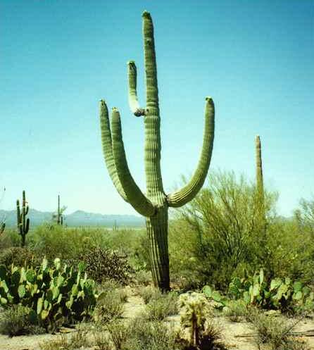
Fig. 13.9. The giant saguaro cactus, Carnegia gigantea, and other large cacti (chollas and prickly pear cacti) are heavily dependent on arbuscular mycorrhzal fungi because these plants have thick, fleshy roots with few or no root hairs. [© Jim Deacon]
Fig. 13.10. Section through part of the protocorm (basal stem region) of an orchid, Neottia, showing coils of hyphae (termed 'peletons') within the orchid cells.The plant cells were alive at the time of sectioning, evidenced by presence of nuclei (the dark granular structures) in two of the orchid cells. [© Jim Deacon] |
||||||||||||