..
MORE IMAGES FROM CHAPTER
10:
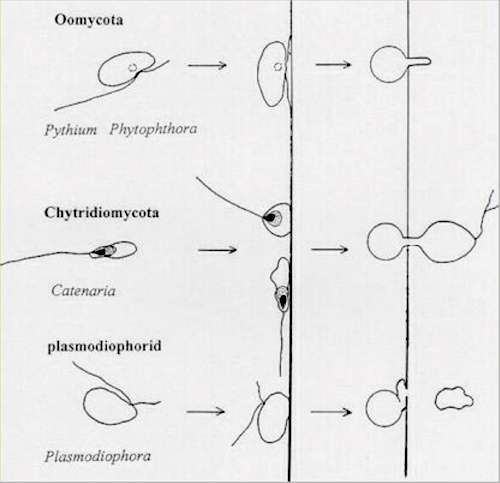
Fig 10.22 Comparison of the docking and penetration of a host by three types of zoosporic fungus (see text for details). In all three types of organism the zoospore shows precise orientation during encystment on a host surface, related to the fact that these zoospores have a pre-determined site from which they penetrate the host. [© Jim Deacon]
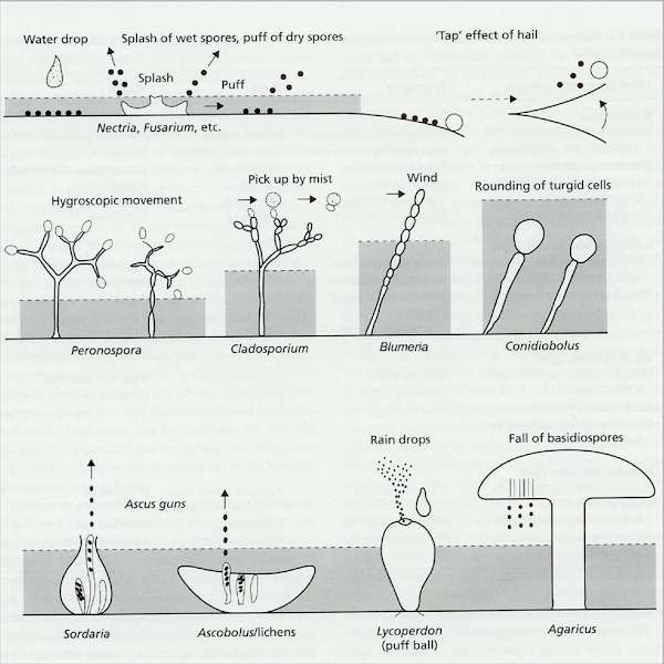
Fig 10.23 The diversity of mechanisms of spore liberation through a boundary layer of still air (shown by shading). [© Jim Deacon]
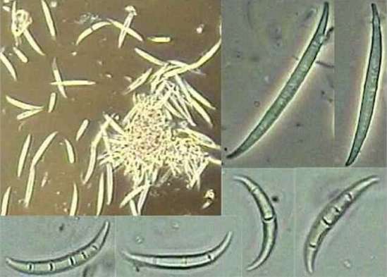
Fig 10.24 Curved spores (macroconidia) with several compartments, typical of many species of Fusarium and other splash-dispersed fungi. [© Jim Deacon]
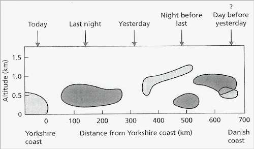
Fig 10.25 Positions of peak spore concentrations of Cladosporium spp. (light shading) and damp-air spore types (dark shading) at different altitudes over the North Sea and at different distances from the English coast. [From Lacey, 1988. based on the work of P. H. Gregory and J. L. Monteith]
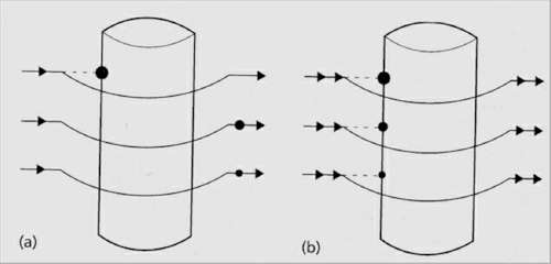
Fig 10.26 Illustration of the relationship between spore size, wind speed and impaction of spores onto a cylinder. (a) At low wind speeds only the largest (heaviest) spores impact. (b) At higher wind speeds progressively smaller (lighter) spores impact. [© Jim Deacon]
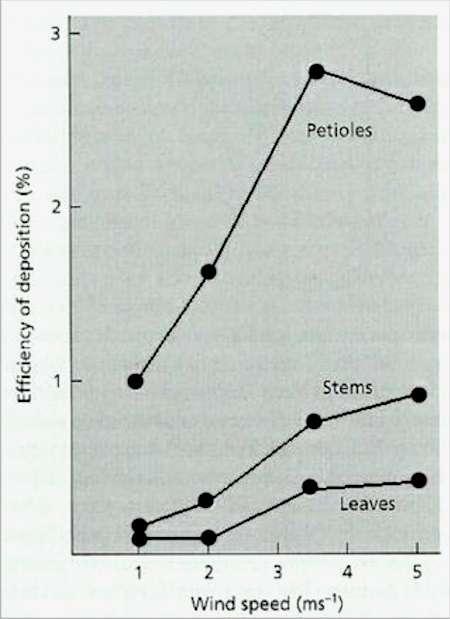
Fig 10.27 Efficiency of impaction of ascospore tetrads of Eutypa armeniacae on leaf blades, petioles, and branch stems of apricot shoots, at different wind speeds. Most impaction occurs on the narrower objects (petioles) and in all cases the efficiency of impaction is increased as the wind speed increases. [From Carter. 1965]
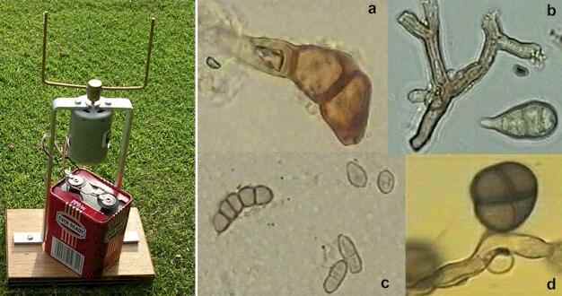
Fig 10.28 The Rotorod air sampler, and some representative spores and hyphal fragments commonly seen when rotorod tapes are examined. Images are at various magnifications: (a) Uredospore of Puccinia graminis. (b) Darkly pigmented hyphae of Cladosporium, with a spore of Alternaria. (c) Various pigmented spores, including Cladosporium (in the lower part of the frame). (d) An immature spore of Epicoccum purpurascens attached to a hyphal fragment [© Jim Deacon]
Fig 10.29 The Burkard continuous monitoring spore sampler. [© Jim Deacon]
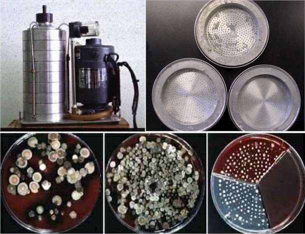
Fig 10.30 Top row: The Anderson spore sampler in assembled form, and three of the metal disks with perforations in their bases. Bottom row: Examples of colonies from agar plates at different levels in an Anderson sampler. [© Jim Deacon]
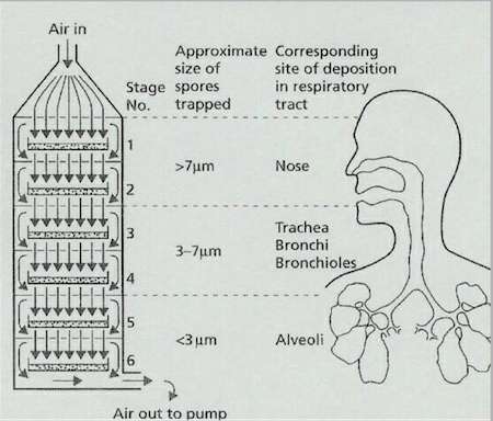
Fig 10.31 Representation of the Anderson sampler, which simulates the deposition of different-sized particles in the human respiratory tract. [© Jim Deacon] |
|||||||||||